


Crystal structure of transketolase from Geobacillus stearothermophilus
Leogrande, C., Rabe von Pappenheim, F., Tittman, K.To be published.
Experimental Data Snapshot
Starting Model: in silico
View more details
Entity ID: 1 | |||||
|---|---|---|---|---|---|
| Molecule | Chains | Sequence Length | Organism | Details | Image |
| Transketolase | 681 | Geobacillus stearothermophilus | Mutation(s): 0 Gene Names: tkt EC: 2.2.1.1 | 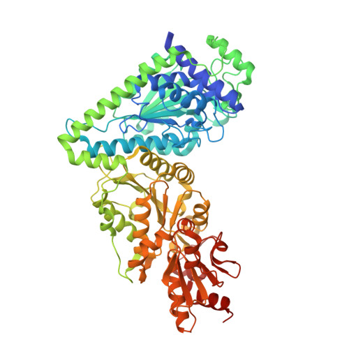 | |
UniProt | |||||
Find proteins for A0A0I9QGZ2 (Geobacillus stearothermophilus) Explore A0A0I9QGZ2 Go to UniProtKB: A0A0I9QGZ2 | |||||
Entity Groups | |||||
| Sequence Clusters | 30% Identity50% Identity70% Identity90% Identity95% Identity100% Identity | ||||
| UniProt Group | A0A0I9QGZ2 | ||||
Sequence AnnotationsExpand | |||||
Reference Sequence | |||||
| Ligands 5 Unique | |||||
|---|---|---|---|---|---|
| ID | Chains | Name / Formula / InChI Key | 2D Diagram | 3D Interactions | |
| TPP (Subject of Investigation/LOI) Download:Ideal Coordinates CCD File | FA [auth C], LA [auth D], M [auth A], U [auth B] | THIAMINE DIPHOSPHATE C12 H19 N4 O7 P2 S AYEKOFBPNLCAJY-UHFFFAOYSA-O |  | ||
| EDO Download:Ideal Coordinates CCD File | AA [auth C] BA [auth C] CA [auth C] E [auth A] EA [auth C] | 1,2-ETHANEDIOL C2 H6 O2 LYCAIKOWRPUZTN-UHFFFAOYSA-N |  | ||
| ACT Download:Ideal Coordinates CCD File | DA [auth C], KA [auth D], L [auth A] | ACETATE ION C2 H3 O2 QTBSBXVTEAMEQO-UHFFFAOYSA-M |  | ||
| CL Download:Ideal Coordinates CCD File | HA [auth C], O [auth A], P [auth A], Q [auth A], W [auth B] | CHLORIDE ION Cl VEXZGXHMUGYJMC-UHFFFAOYSA-M |  | ||
| MG Download:Ideal Coordinates CCD File | GA [auth C], MA [auth D], N [auth A], V [auth B] | MAGNESIUM ION Mg JLVVSXFLKOJNIY-UHFFFAOYSA-N |  | ||
| Length ( Å ) | Angle ( ˚ ) |
|---|---|
| a = 79.44 | α = 80.89 |
| b = 82.71 | β = 68.34 |
| c = 106.14 | γ = 69.99 |
| Software Name | Purpose |
|---|---|
| PHENIX | refinement |
| XDS | data reduction |
| XSCALE | data scaling |
| PHENIX | phasing |
| Funding Organization | Location | Grant Number |
|---|---|---|
| H2020 Marie Curie Actions of the European Commission | European Union | 956631 |


