8CIC

Crystal structure of stabilized A2A adenosine receptor A2AR-StaR2-bRIL in complex with clinical candidate Etrumadenant


Experimental Data Snapshot

  • Method: X-RAY DIFFRACTION
  • Resolution: 2.10 Å
  • R-Value Free: 0.214 
  • R-Value Work: 0.190 
  • R-Value Observed: 0.192 

wwPDB Validation   3D Report Full Report


Ligand Structure Quality Assessment 


This is version 1.1 of the entry. See complete history


Literature

Crystal structure of adenosine A 2A receptor in complex with clinical candidate Etrumadenant reveals unprecedented antagonist interaction.

Claff, T.Schlegel, J.G.Voss, J.H.Vaassen, V.J.Weisse, R.H.Cheng, R.K.Y.Markovic-Mueller, S.Bucher, D.Strater, N.Muller, C.E.

(2023) Commun Chem 6: 106-106

  • DOI: https://doi.org/10.1038/s42004-023-00894-6
  • Primary Citation of Related Structures:  
    8C9W, 8CIC

  • PubMed Abstract: 

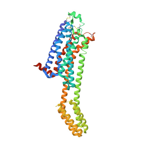
    The G s  protein-coupled adenosine A 2A receptor (A 2A AR) represents an emerging drug target for cancer immunotherapy. The clinical candidate Etrumadenant was developed as an A 2A AR antagonist with ancillary blockade of the A 2B AR subtype. It constitutes a unique chemotype featuring a poly-substituted 2-amino-4-phenyl-6-triazolylpyrimidine core structure. Herein, we report two crystal structures of the A 2A AR in complex with Etrumadenant, obtained with differently thermostabilized A 2A AR constructs. This led to the discovery of an unprecedented interaction, a hydrogen bond of T88 3.36 with the cyano group of Etrumadenant. T88 3.36 is mutated in most A 2A AR constructs used for crystallization, which has prevented the discovery of its interactions. In-vitro characterization of Etrumadenant indicated low selectivity versus the A 1 AR subtype, which can be rationalized by the structural data. These results will facilitate the future design of AR antagonists with desired selectivity. Moreover, they highlight the advantages of the employed A 2A AR crystallization construct that is devoid of ligand binding site mutations.


  • Organizational Affiliation

    PharmaCenter Bonn & Pharmaceutical Institute, Department of Pharmaceutical & Medicinal Chemistry, University of Bonn, An der Immenburg 4, 53113, Bonn, Germany. tobias.claff@uni-bonn.de.


Macromolecules
Find similar proteins by:  (by identity cutoff)  |  3D Structure
Entity ID: 1
MoleculeChains Sequence LengthOrganismDetailsImage
Adenosine receptor A2a,Soluble cytochrome b562433Homo sapiensMutation(s): 11 
Gene Names: ADORA2AADORA2cybC
Membrane Entity: Yes 
UniProt & NIH Common Fund Data Resources
Find proteins for P0ABE7 (Escherichia coli)
Explore P0ABE7 
Go to UniProtKB:  P0ABE7
Find proteins for P29274 (Homo sapiens)
Explore P29274 
Go to UniProtKB:  P29274
PHAROS:  P29274
GTEx:  ENSG00000128271 
Entity Groups  
Sequence Clusters30% Identity50% Identity70% Identity90% Identity95% Identity100% Identity
UniProt GroupsP0ABE7P29274
Sequence Annotations
Expand
  • Reference Sequence
Small Molecules
Ligands 6 Unique
IDChains Name / Formula / InChI Key2D Diagram3D Interactions
U30 (Subject of Investigation/LOI)
Query on U30

Download Ideal Coordinates CCD File 
C [auth A]3-[2-azanyl-6-[1-[[6-(2-oxidanylpropan-2-yl)pyridin-2-yl]methyl]-1,2,3-triazol-4-yl]pyrimidin-4-yl]-2-methyl-benzenecarbonitrile
C23 H22 N8 O
BUXIAWLTBSXYSW-UHFFFAOYSA-N
CLR
Query on CLR

Download Ideal Coordinates CCD File 
D [auth A],
E [auth A],
F [auth A],
G [auth A]
CHOLESTEROL
C27 H46 O
HVYWMOMLDIMFJA-DPAQBDIFSA-N
OLC
Query on OLC

Download Ideal Coordinates CCD File 
AA [auth A],
BA [auth A],
CA [auth A],
DA [auth A],
Z [auth A]
(2R)-2,3-dihydroxypropyl (9Z)-octadec-9-enoate
C21 H40 O4
RZRNAYUHWVFMIP-GDCKJWNLSA-N
OLB
Query on OLB

Download Ideal Coordinates CCD File 
W [auth A],
X [auth A],
Y [auth A]
(2S)-2,3-dihydroxypropyl (9Z)-octadec-9-enoate
C21 H40 O4
RZRNAYUHWVFMIP-QJRAZLAKSA-N
OLA
Query on OLA

Download Ideal Coordinates CCD File 
H [auth A]
I [auth A]
J [auth A]
K [auth A]
L [auth A]
H [auth A],
I [auth A],
J [auth A],
K [auth A],
L [auth A],
M [auth A],
N [auth A],
O [auth A],
P [auth A],
Q [auth A],
R [auth A],
S [auth A],
T [auth A],
U [auth A],
V [auth A]
OLEIC ACID
C18 H34 O2
ZQPPMHVWECSIRJ-KTKRTIGZSA-N
NA
Query on NA

Download Ideal Coordinates CCD File 
B [auth A]SODIUM ION
Na
FKNQFGJONOIPTF-UHFFFAOYSA-N
Experimental Data & Validation

Experimental Data

  • Method: X-RAY DIFFRACTION
  • Resolution: 2.10 Å
  • R-Value Free: 0.214 
  • R-Value Work: 0.190 
  • R-Value Observed: 0.192 
  • Space Group: C 2 2 21
Unit Cell:
Length ( Å )Angle ( ˚ )
a = 39.36α = 90
b = 179.091β = 90
c = 140.573γ = 90
Software Package:
Software NamePurpose
PHENIXrefinement
Aimlessdata scaling
XDSdata reduction
PHASERphasing

Structure Validation

View Full Validation Report


Ligand Structure Quality Assessment 


Entry History & Funding Information

Deposition Data


Funding OrganizationLocationGrant Number
Not funded--

Revision History  (Full details and data files)

  • Version 1.0: 2023-05-31
    Type: Initial release
  • Version 1.1: 2023-06-14
    Changes: Database references