8C0V

Structure of the peroxisomal Pex1/Pex6 ATPase complex bound to a substrate in single seam state


Experimental Data Snapshot

  • Method: ELECTRON MICROSCOPY
  • Resolution: 4.10 Å
  • Aggregation State: PARTICLE 
  • Reconstruction Method: SINGLE PARTICLE 

wwPDB Validation   3D Report Full Report


This is version 1.0 of the entry. See complete history


Literature

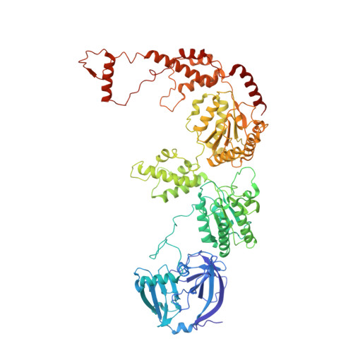
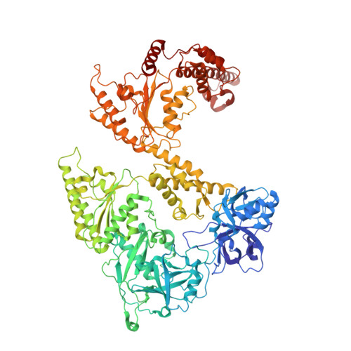
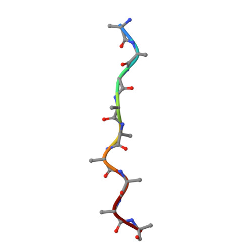
Structure of the peroxisomal Pex1/Pex6 ATPase complex bound to a substrate.

Ruttermann, M.Koci, M.Lill, P.Geladas, E.D.Kaschani, F.Klink, B.U.Erdmann, R.Gatsogiannis, C.

(2023) Nat Commun 14: 5942-5942

  • DOI: https://doi.org/10.1038/s41467-023-41640-9
  • Primary Citation of Related Structures:  
    8C0V, 8C0W

  • PubMed Abstract: 

    The double-ring AAA+ ATPase Pex1/Pex6 is required for peroxisomal receptor recycling and is essential for peroxisome formation. Pex1/Pex6 mutations cause severe peroxisome associated developmental disorders. Despite its pathophysiological importance, mechanistic details of the heterohexamer are not yet available. Here, we report cryoEM structures of Pex1/Pex6 from Saccharomyces cerevisiae, with an endogenous protein substrate trapped in the central pore of the catalytically active second ring (D2). Pairs of Pex1/Pex6(D2) subdomains engage the substrate via a staircase of pore-1 loops with distinct properties. The first ring (D1) is catalytically inactive but undergoes significant conformational changes resulting in alternate widening and narrowing of its pore. These events are fueled by ATP hydrolysis in the D2 ring and disengagement of a "twin-seam" Pex1/Pex6(D2) heterodimer from the staircase. Mechanical forces are propagated in a unique manner along Pex1/Pex6 interfaces that are not available in homo-oligomeric AAA-ATPases. Our structural analysis reveals the mechanisms of how Pex1 and Pex6 coordinate to achieve substrate translocation.


  • Organizational Affiliation

    Institute for Medical Physics and Biophysics, University Münster, Münster, Germany.


Macromolecules
Find similar proteins by:  (by identity cutoff)  |  3D Structure
Entity ID: 1
MoleculeChains Sequence LengthOrganismDetailsImage
Peroxisomal ATPase PEX1
A, C, E
823Saccharomyces cerevisiaeMutation(s): 0 
Gene Names: PEX1PAS1YKL197C
EC: 3.6.4
UniProt
Find proteins for P24004 (Saccharomyces cerevisiae (strain ATCC 204508 / S288c))
Explore P24004 
Go to UniProtKB:  P24004
Entity Groups  
Sequence Clusters30% Identity50% Identity70% Identity90% Identity95% Identity100% Identity
UniProt GroupP24004
Sequence Annotations
Expand
  • Reference Sequence
Find similar proteins by:  (by identity cutoff)  |  3D Structure
Entity ID: 2
MoleculeChains Sequence LengthOrganismDetailsImage
Peroxisomal ATPase PEX6
B, D, F
1,030Saccharomyces cerevisiaeMutation(s): 1 
Gene Names: PEX6PAS8YNL329CN0310
EC: 3.6.4
UniProt
Find proteins for P33760 (Saccharomyces cerevisiae (strain ATCC 204508 / S288c))
Explore P33760 
Go to UniProtKB:  P33760
Entity Groups  
Sequence Clusters30% Identity50% Identity70% Identity90% Identity95% Identity100% Identity
UniProt GroupP33760
Sequence Annotations
Expand
  • Reference Sequence

Find similar proteins by:  Sequence   |   3D Structure  

Entity ID: 3
MoleculeChains Sequence LengthOrganismDetailsImage
unknown peptideG [auth R]9Saccharomyces cerevisiaeMutation(s): 0 
Sequence Annotations
Expand
  • Reference Sequence
Small Molecules
Ligands 3 Unique
IDChains Name / Formula / InChI Key2D Diagram3D Interactions
ATP (Subject of Investigation/LOI)
Query on ATP

Download Ideal Coordinates CCD File 
AA [auth F]
H [auth A]
I [auth A]
L [auth B]
M [auth B]
AA [auth F],
H [auth A],
I [auth A],
L [auth B],
M [auth B],
P [auth C],
Q [auth C],
T [auth D],
U [auth D],
X [auth E]
ADENOSINE-5'-TRIPHOSPHATE
C10 H16 N5 O13 P3
ZKHQWZAMYRWXGA-KQYNXXCUSA-N
ADP (Subject of Investigation/LOI)
Query on ADP

Download Ideal Coordinates CCD File 
BA [auth F],
Y [auth E]
ADENOSINE-5'-DIPHOSPHATE
C10 H15 N5 O10 P2
XTWYTFMLZFPYCI-KQYNXXCUSA-N
MG
Query on MG

Download Ideal Coordinates CCD File 
CA [auth F]
J [auth A]
K [auth A]
N [auth B]
O [auth B]
CA [auth F],
J [auth A],
K [auth A],
N [auth B],
O [auth B],
R [auth C],
S [auth C],
V [auth D],
W [auth D],
Z [auth E]
MAGNESIUM ION
Mg
JLVVSXFLKOJNIY-UHFFFAOYSA-N
Experimental Data & Validation

Experimental Data

  • Method: ELECTRON MICROSCOPY
  • Resolution: 4.10 Å
  • Aggregation State: PARTICLE 
  • Reconstruction Method: SINGLE PARTICLE 
EM Software:
TaskSoftware PackageVersion
RECONSTRUCTIONRELION3.1.4
MODEL REFINEMENTPHENIX1.2

Structure Validation

View Full Validation Report


Entry History & Funding Information

Deposition Data


Funding OrganizationLocationGrant Number
German Research Foundation (DFG)GermanyGA 2519/1-2
German Research Foundation (DFG)GermanyER 178/7-2
German Research Foundation (DFG)GermanyINST 211/667-1
German Research Foundation (DFG)GermanySFB 1430/1, A04

Revision History  (Full details and data files)

  • Version 1.0: 2023-10-04
    Type: Initial release