8BLZ

Engineered Fructosyl Peptide Oxidase - D02 mutant


Experimental Data Snapshot

  • Method: X-RAY DIFFRACTION
  • Resolution: 1.70 Å
  • R-Value Free: 0.194 
  • R-Value Work: 0.168 
  • R-Value Observed: 0.169 

wwPDB Validation   3D Report Full Report


Ligand Structure Quality Assessment 


This is version 1.1 of the entry. See complete history


Literature

Tailoring FPOX enzymes for enhanced stability and expanded substrate recognition.

Estiri, H.Bhattacharya, S.Buitrago, J.A.R.Castagna, R.Legzdina, L.Casucci, G.Ricci, A.Parisini, E.Gautieri, A.

(2023) Sci Rep 13: 18610-18610

  • DOI: https://doi.org/10.1038/s41598-023-45428-1
  • Primary Citation of Related Structures:  
    8BJY, 8BLX, 8BLZ, 8BMU

  • PubMed Abstract: 

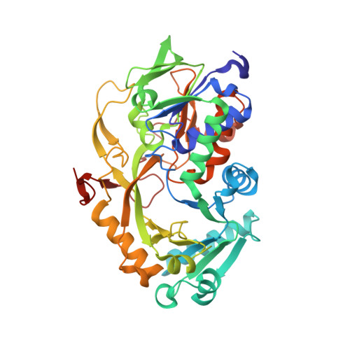
    Fructosyl peptide oxidases (FPOX) are deglycating enzymes that find application as key enzymatic components in diabetes monitoring devices. Indeed, their use with blood samples can provide a measurement of the concentration of glycated hemoglobin and glycated albumin, two well-known diabetes markers. However, the FPOX currently employed in enzymatic assays cannot directly detect whole glycated proteins, making it necessary to perform a preliminary proteolytic treatment of the target protein to generate small glycated peptides that can act as viable substrates for the enzyme. This is a costly and time consuming step. In this work, we used an in silico protein engineering approach to enhance the overall thermal stability of the enzyme and to improve its catalytic activity toward large substrates. The final design shows a marked improvement in thermal stability relative to the wild type enzyme, a distinct widening of its access tunnel and significant enzymatic activity towards a range of glycated substrates.


  • Organizational Affiliation

    Department of Biotechnology, Latvian Institute of Organic Synthesis, Aizkraukles 21, Riga, 1006, Latvia.


Macromolecules
Find similar proteins by:  (by identity cutoff)  |  3D Structure
Entity ID: 1
MoleculeChains Sequence LengthOrganismDetailsImage
Fructosyl Peptide Oxidase mutant (D02)A [auth C]424Parastagonospora nodorum SN15Mutation(s): 0 
Gene Names: SNOG_08398
UniProt
Find proteins for Q0UIL6 (Phaeosphaeria nodorum (strain SN15 / ATCC MYA-4574 / FGSC 10173))
Explore Q0UIL6 
Go to UniProtKB:  Q0UIL6
Entity Groups  
Sequence Clusters30% Identity50% Identity70% Identity90% Identity95% Identity100% Identity
UniProt GroupQ0UIL6
Sequence Annotations
Expand
  • Reference Sequence
Small Molecules
Ligands 3 Unique
IDChains Name / Formula / InChI Key2D Diagram3D Interactions
FAD (Subject of Investigation/LOI)
Query on FAD

Download Ideal Coordinates CCD File 
B [auth C]FLAVIN-ADENINE DINUCLEOTIDE
C27 H33 N9 O15 P2
VWWQXMAJTJZDQX-UYBVJOGSSA-N
SO4
Query on SO4

Download Ideal Coordinates CCD File 
I [auth C],
J [auth C]
SULFATE ION
O4 S
QAOWNCQODCNURD-UHFFFAOYSA-L
GOL
Query on GOL

Download Ideal Coordinates CCD File 
C
D [auth C]
E [auth C]
F [auth C]
G [auth C]
C,
D [auth C],
E [auth C],
F [auth C],
G [auth C],
H [auth C]
GLYCEROL
C3 H8 O3
PEDCQBHIVMGVHV-UHFFFAOYSA-N
Experimental Data & Validation

Experimental Data

  • Method: X-RAY DIFFRACTION
  • Resolution: 1.70 Å
  • R-Value Free: 0.194 
  • R-Value Work: 0.168 
  • R-Value Observed: 0.169 
  • Space Group: C 2 2 21
Unit Cell:
Length ( Å )Angle ( ˚ )
a = 91.096α = 90
b = 129.493β = 90
c = 86.78γ = 90
Software Package:
Software NamePurpose
REFMACrefinement
DIALSdata reduction
Aimlessdata scaling
Cootmodel building
MOLREPphasing

Structure Validation

View Full Validation Report


Ligand Structure Quality Assessment 


Entry History & Funding Information

Deposition Data


Funding OrganizationLocationGrant Number
Other governmentlzp-2020/2-0013

Revision History  (Full details and data files)

  • Version 1.0: 2023-11-22
    Type: Initial release
  • Version 1.1: 2024-06-05
    Changes: Database references