Serial Femtosecond Crystallography Reveals that Photoactivation in a Fluorescent Protein Proceeds via the Hula Twist Mechanism.
Fadini, A., Hutchison, C.D.M., Morozov, D., Chang, J., Maghlaoui, K., Perrett, S., Luo, F., Kho, J.C.X., Romei, M.G., Morgan, R.M.L., Orr, C.M., Cordon-Preciado, V., Fujiwara, T., Nuemket, N., Tosha, T., Tanaka, R., Owada, S., Tono, K., Iwata, S., Boxer, S.G., Groenhof, G., Nango, E., van Thor, J.J.(2023) J Am Chem Soc
- PubMed: 37418747 Search on PubMedSearch on PubMed Central
- DOI: https://doi.org/10.1021/jacs.3c02313
- Primary Citation Related Structures:
8A6G, 8A6N, 8A6O, 8A6P, 8A6Q, 8A6R, 8A6S, 8A7V, 8A83, 8AM4 - PubMed Abstract:
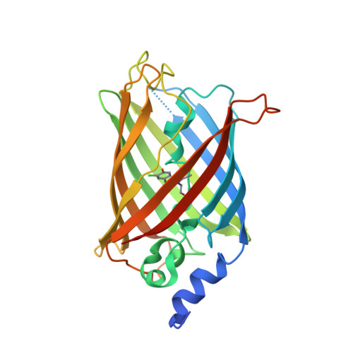
Chromophore cis/trans photoisomerization is a fundamental process in chemistry and in the activation of many photosensitive proteins. A major task is understanding the effect of the protein environment on the efficiency and direction of this reaction compared to what is observed in the gas and solution phases. In this study, we set out to visualize the hula twist (HT) mechanism in a fluorescent protein, which is hypothesized to be the preferred mechanism in a spatially constrained binding pocket. We use a chlorine substituent to break the twofold symmetry of the embedded phenolic group of the chromophore and unambiguously identify the HT primary photoproduct. Through serial femtosecond crystallography, we then track the photoreaction from femtoseconds to the microsecond regime. We observe signals for the photoisomerization of the chromophore as early as 300 fs, obtaining the first experimental structural evidence of the HT mechanism in a protein on its femtosecond-to-picosecond timescale. We are then able to follow how chromophore isomerization and twisting lead to secondary structure rearrangements of the protein β-barrel across the time window of our measurements.
- Department of Life Sciences, Faculty of Natural Sciences, Imperial College London, London SW7 2AZ, U.K.
Organizational Affiliation: