Targeting the phosphoserine phosphatase MtSerB2 for tuberculosis drug discovery, an hybrid knowledge based /fragment based approach.
Haufroid, M., Volkov, A.N., Wouters, J.(2022) Eur J Med Chem 245: 114935-114935
- PubMed: 36403421 Search on PubMed
- DOI: https://doi.org/10.1016/j.ejmech.2022.114935
- Primary Citation Related Structures:
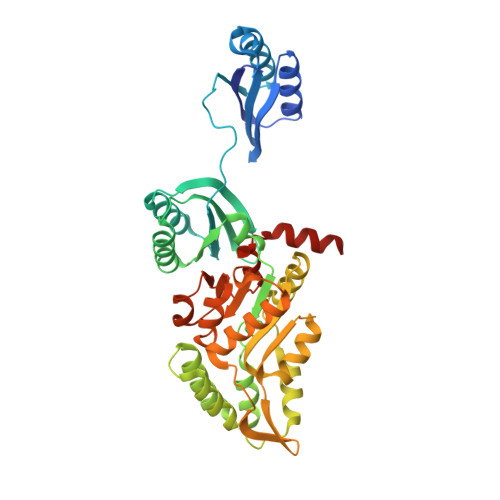
8A1Z, 8A21 - PubMed Abstract:
Tuberculosis is currently still one of the leading causes of death from a treatable pathogen. The proportion of cases of resistance to common antibiotics is frequently increasing and the development of new drugs with new therapeutic targets is becoming necessary. The Mycobacterium tuberculosis phosphoserine phosphatase MtSerB2 is an interesting enzyme to target in drug design because of its ability to allow immune evasion of the bacteria. Research has already been carried out on this protein both from a mechanistic point of view and from the point of view of its inhibition by trisubstituted harmine derivatives. Based on this work, a new approach based on virtual screening is presented in the selection of fragment-sized harmine-derived compounds as well as chelators to target the catalytic magnesium of MtSerB2. The selection of a minimum list of fragments is explained as well as the screening cascade (DSF, Ligand-based NMR, High concentration enzymatic assay) to characterise their affinity for MtSerB2. Crystallogenesis assays have provided structural information on some promising fragments and the development of a pharmacophore model with the structural elements necessary for the development of more complex inhibitors. Ultimately, this work on fragment growth would allow the development of antimycobacterial molecules inhibiting MtSerB2 as well as the growth of the pathogen.
- Namur Medicine and Drug Innovation Center, Namur Research Institute for Life Science NAMEDIC-NARILIS, Department of Chemistry, Laboratoire de Chimie Biologique Structurale (CBS), University of Namur (UNamur), Rue de Bruxelles 61, 5000, Namur, Belgium. Electronic address: marie.haufroid@unamur.be.
Organizational Affiliation: