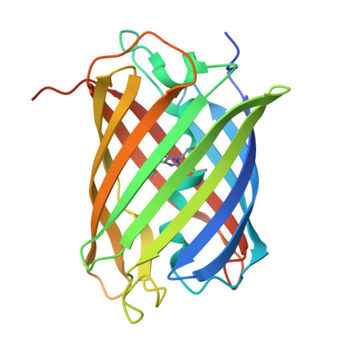
The stability of fluorescent proteins (FPs) is crucial for imaging techniques such as live-cell imaging, super-resolution microscopy and correlative light and electron microscopy. Although stable green and yellow FPs are available, stable monomeric red FPs (RFPs) remain limited. Here we develop an extremely stable monomeric RFP named mScarlet3-H and determine its structure at a 1.5 Å resolution. mScarlet3-H exhibits remarkable resistance to high temperature, chaotropic conditions and oxidative environments, enabling efficient correlative light and electron microscopy imaging and rapid (less than 1 day) whole-organ tissue clearing. In addition, its high photostability allows long-term three-dimensional structured illumination microscopy imaging of mitochondrial dynamics with minimal photobleaching. It also facilitates dual-color live-cell stimulated emission depletion imaging with a high signal-to-noise ratio and strong specificity. Systematic benchmarking against high-performing RFPs established mScarlet3-H as a highly stable RFP for multimodality microscopy in cell cultures and model organisms, complementing green FPs for multiplexed imaging in zebrafish, mice and Nicotiana benthamiana.
Organizational Affiliation:
Key Laboratory of Clinical Laboratory Technology for Precision Medicine, Institute of Neuroscience, and Fujian Key Laboratory of Molecular Neurology, Public Technology Service Center, Fujian Medical University, Fuzhou, China.
The School of Basic Medical Sciences, Fujian Medical University, Fuzhou, China.
Academy for Advanced Interdisciplinary Studies, Peking University, Beijing, China.
Chinese Institute for Brain Research, Beijing, China.
School of Life Sciences, Westlake University, Hangzhou, China.
Westlake Laboratory of Life Sciences and Biomedicine, Hangzhou, China.
Institute of Basic Medical Sciences, Westlake Institute for Advanced Study, Hangzhou, China.
College of Life Sciences, Zhejiang University, Hangzhou, China.
State Key Laboratory of Vaccines for Infectious Diseases, National Institute of Diagnostics and Vaccine Development in Infectious Diseases, School of Public Health, Xiamen University, Xiamen, China.
Microscopy core facility of Westlake University, Hangzhou, China.
Institute of Life Sciences, Fuzhou University, Fuzhou, China.
National Laboratory of Biomacromolecules, New Cornerstone Science Laboratory, CAS Center for Excellence in Biomacromolecules, Institute of Biophysics, Chinese Academy of Sciences, Beijing, China.
Key Laboratory of Ministry of Education for Gastrointestinal Cancer, School of Basic Medical Sciences, Fujian Medical University, Fuzhou, China.
Fujian Key Laboratory of Drug Target Discovery and Structural and Functional Research, School of Pharmacy, Fujian Medical University, Fuzhou, China.
School of Pharmacy, Center of Translational Hematology, Fujian Medical University, Fuzhou, China.
State Key Laboratory of Cellular Stress Biology, School of Life Sciences, Faculty of Medicine and Life Sciences, Xiamen University, Xiamen, China.
X-ray crystallography platform of National Protein Science Facility, Tsinghua University, Beijing, China.
Key Laboratory of Clinical Laboratory Technology for Precision Medicine, Institute of Neuroscience, and Fujian Key Laboratory of Molecular Neurology, Public Technology Service Center, Fujian Medical University, Fuzhou, China. congxianwu@fjmu.edu.cn.
State Key Laboratory of Vaccines for Infectious Diseases, National Institute of Diagnostics and Vaccine Development in Infectious Diseases, School of Public Health, Xiamen University, Xiamen, China. abing0811@xmu.edu.cn.
Westlake Laboratory of Life Sciences and Biomedicine, Hangzhou, China. kiryl.piatkevich@westlake.edu.cn.
Institute of Basic Medical Sciences, Westlake Institute for Advanced Study, Hangzhou, China. kiryl.piatkevich@westlake.edu.cn.
College of Life Sciences, Zhejiang University, Hangzhou, China. kiryl.piatkevich@westlake.edu.cn.
Key Laboratory of Clinical Laboratory Technology for Precision Medicine, Institute of Neuroscience, and Fujian Key Laboratory of Molecular Neurology, Public Technology Service Center, Fujian Medical University, Fuzhou, China. fuzhifei@fjmu.edu.cn.