8ZUS | pdb_00008zus

Crystal structure of the F99S/M153T/V163A/T203V variant of GFP at pH 7.5


Experimental Data Snapshot

  • Method: X-RAY DIFFRACTION
  • Resolution: 1.20 Å
  • R-Value Free: 
    0.165 (Depositor), 0.165 (DCC) 
  • R-Value Work: 
    0.133 (Depositor), 0.133 (DCC) 
  • R-Value Observed: 
    0.134 (Depositor) 

Starting Model: experimental
View more details

wwPDB Validation 3D Report Full Report

Validation slider image for 8ZUS

This is version 2.0 of the entry. See complete history

Literature

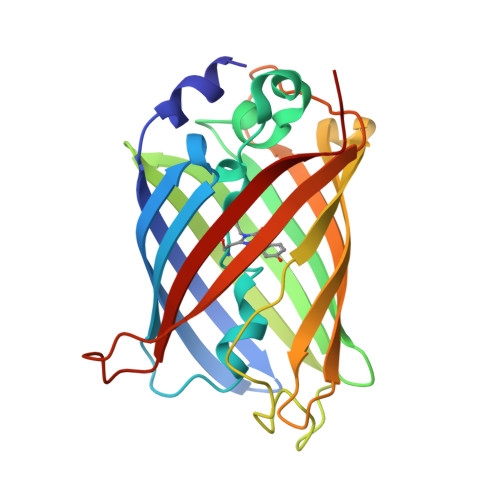
Structural characterization of green fluorescent protein in the I-state.

Takeda, R.Tsutsumi, E.Okatsu, K.Fukai, S.Takeda, K.

(2024) Sci Rep 14: 22832-22832

  • DOI: https://doi.org/10.1038/s41598-024-73696-y
  • Primary Citation Related Structures: 
    8ZUP, 8ZUQ, 8ZUR, 8ZUS, 8ZUT

  • PubMed Abstract: 

    Green fluorescent protein (GFP) is widely utilized as a fluorescent tag in biochemical fields. Whereas the intermediate (I) state has been proposed in the photoreaction cycle in addition to the A and B states, until now the structure of I has only been estimated by computational studies. In this paper, we report the crystal structures of the I stabilizing variants of GFP at high resolutions where respective atoms can be observed separately. Comparison with the structures in the other states highlights the structural feature of the I state. The side chain of one of the substituted residues, Val203, adopts the gauche- conformation observed for Thr203 in the A state, which is different from the B state. On the other hand, His148 interacts with the chromophore by ordinary hydrogen bonding with a distance of 2.85 Å, while the weaker interaction by longer distances is observed in the A state. Therefore, it was indicated that it is possible to distinguish three states A, B and I by the two hydrogen bond distances Oγ-Thr203···Oη-chromophore and Nδ1-His148···Oη-chromophore. We discuss the characteristics of the I intermediate of wild-type GFP on the bases of the structure estimated from the variant structures by quantum chemical calculations.


  • Organizational Affiliation
    • Department of Chemistry, Graduate School of Science, Kyoto University, Sakyo-ku, Kyoto, 606-8502, Japan.

Macromolecule Content 

  • Total Structure Weight: 25.89 kDa 
  • Atom Count: 2,714 
  • Modeled Residue Count: 228 
  • Deposited Residue Count: 228 
  • Unique protein chains: 1

Macromolecules

Find similar proteins by:|  3D Structure
Entity ID: 1
MoleculeChains  Sequence LengthOrganismDetailsImage
Green fluorescent protein228Aequorea victoriaMutation(s): 5 
Gene Names: GFP
UniProt
Find proteins for P42212 (Aequorea victoria)
Explore P42212 
Go to UniProtKB:  P42212
Entity Groups
Sequence Clusters30% Identity50% Identity70% Identity90% Identity95% Identity100% Identity
UniProt GroupP42212
Sequence Annotations
Expand
Reference Sequence

Small Molecules

Modified Residues  1 Unique
IDChains TypeFormula2D DiagramParent
GYS
Query on GYS
A
L-PEPTIDE LINKINGC14 H15 N3 O5SER, TYR, GLY

Experimental Data & Validation

Experimental Data

  • Method: X-RAY DIFFRACTION
  • Resolution: 1.20 Å
  • R-Value Free:  0.165 (Depositor), 0.165 (DCC) 
  • R-Value Work:  0.133 (Depositor), 0.133 (DCC) 
  • R-Value Observed: 0.134 (Depositor) 
Space Group: P 21 21 21
Unit Cell:
Length ( Å )Angle ( ˚ )
a = 50.72α = 90
b = 62.236β = 90
c = 68.343γ = 90
Software Package:
Software NamePurpose
PHENIXrefinement
XDSdata scaling
XDSdata reduction
PHENIXphasing

Structure Validation

View Full Validation Report


Entry History 

& Funding Information

Deposition Data


Funding OrganizationLocationGrant Number
Takeda Science FoundationJapan--
Gimpu fund from Kyoto UniversityJapan--

Revision History  (Full details and data files)

  • Version 1.0: 2024-11-06
    Type: Initial release
  • Version 1.1: 2025-06-18
    Changes: Database references
  • Version 2.0: 2026-03-18
    Changes: Polymer sequence