8YM2 | pdb_00008ym2

Crystal structure of AIDA-1 PTB domain in complex with SynGAP NPxF motif


Experimental Data Snapshot

  • Method: X-RAY DIFFRACTION
  • Resolution: 2.00 Å
  • R-Value Free: 
    0.264 (Depositor), 0.266 (DCC) 
  • R-Value Work: 
    0.213 (Depositor), 0.222 (DCC) 
  • R-Value Observed: 
    0.216 (Depositor) 

Starting Model: experimental
View more details

wwPDB Validation 3D Report Full Report

Validation slider image for 8YM2

This is version 1.1 of the entry. See complete history

Literature

AIDA-1/ANKS1B Binds to the SynGAP Family RasGAPs with High Affinity and Specificity.

Wang, X.Wang, Y.Cai, Q.Zhang, M.

(2024) J Mol Biology 436: 168608-168608

  • DOI: https://doi.org/10.1016/j.jmb.2024.168608
  • Primary Citation Related Structures: 
    8YM2

  • PubMed Abstract: 

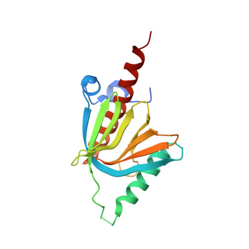
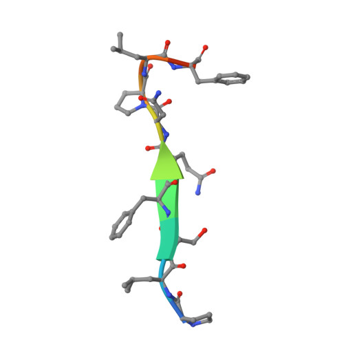
    AIDA-1, encoded by ANKS1B, is an abundant postsynaptic scaffold protein essential for brain development. Mutations of ANKS1B are closely associated with various psychiatric disorders. However, very little is known regarding the molecular mechanisms underlying AIDA-1's involvements under physiological and pathophysiological conditions. Here, we discovered an interaction between AIDA-1 and the SynGAP family Ras-GTPase activating protein (GAP) via affinity purification using AIDA-1d as the bait. Biochemical studies showed that the PTB domain of AIDA-1 binds to an extended NPx[F/Y]-motif of the SynGAP family proteins with high affinities. The high-resolution crystal structure of AIDA-1 PTB domain in complex with the SynGAP NPxF-motif revealed the molecular mechanism governing the specific interaction between AIDA-1 and SynGAP. Our study not only explains why patients with ANKS1B or SYNGAP1 mutations share overlapping clinical phenotypes, but also allows identification of new AIDA-1 binding targets such as Ras and Rab interactors.


  • Organizational Affiliation
    • Biomedical Research Institute, Shenzhen Peking University-The Hong Kong University of Science and Technology Medical Center, Shenzhen 518036, China. Electronic address: xueqwang@pku.edu.cn.

Macromolecule Content 

  • Total Structure Weight: 19.58 kDa 
  • Atom Count: 1,295 
  • Modeled Residue Count: 158 
  • Deposited Residue Count: 173 
  • Unique protein chains: 2

Macromolecules

Find similar proteins by:|  3D Structure
Entity ID: 1
MoleculeChains  Sequence LengthOrganismDetailsImage
Ankyrin repeat and sterile alpha motif domain-containing protein 1B162Mus musculusMutation(s): 0 
Gene Names: Anks1b
UniProt
Find proteins for Q8BIZ1 (Mus musculus)
Explore Q8BIZ1 
Go to UniProtKB:  Q8BIZ1
Entity Groups
Sequence Clusters30% Identity50% Identity70% Identity90% Identity95% Identity100% Identity
UniProt GroupQ8BIZ1
Sequence Annotations
Expand
Reference Sequence
Find similar proteins by:  Sequence   |   3D Structure  
Entity ID: 2
MoleculeChains  Sequence LengthOrganismDetailsImage
Ras/Rap GTPase-activating protein SynGAP11Rattus norvegicusMutation(s): 0 
UniProt
Find proteins for Q9QUH6 (Rattus norvegicus)
Explore Q9QUH6 
Go to UniProtKB:  Q9QUH6
Entity Groups
Sequence Clusters30% Identity50% Identity70% Identity90% Identity95% Identity100% Identity
UniProt GroupQ9QUH6
Sequence Annotations
Expand
Reference Sequence

Experimental Data & Validation

Experimental Data

  • Method: X-RAY DIFFRACTION
  • Resolution: 2.00 Å
  • R-Value Free:  0.264 (Depositor), 0.266 (DCC) 
  • R-Value Work:  0.213 (Depositor), 0.222 (DCC) 
  • R-Value Observed: 0.216 (Depositor) 
Space Group: I 2 3
Unit Cell:
Length ( Å )Angle ( ˚ )
a = 112.919α = 90
b = 112.919β = 90
c = 112.919γ = 90
Software Package:
Software NamePurpose
REFMACrefinement
HKL-2000data scaling
HKL-2000data reduction
PHASERphasing

Structure Validation

View Full Validation Report


Entry History 

& Funding Information

Deposition Data


Funding OrganizationLocationGrant Number
Ministry of Science and Technology (MoST, China)China2019YFA0508402

Revision History  (Full details and data files)

  • Version 1.0: 2024-05-29
    Type: Initial release
  • Version 1.1: 2024-06-05
    Changes: Database references