Structural and biochemical analysis of penicillin-binding protein 2 from Campylobacter jejuni.
Choi, H.J., Ki, D.U., Yoon, S.I.(2024) Biochem Biophys Res Commun 710: 149859-149859
- PubMed: 38581948 Search on PubMed
- DOI: https://doi.org/10.1016/j.bbrc.2024.149859
- Primary Citation Related Structures:
8YJX - PubMed Abstract:
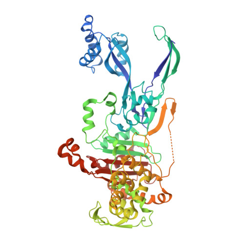
Penicillin-binding protein 2 (PBP2) plays a key role in the formation of peptidoglycans in bacterial cell walls by crosslinking glycan chains through transpeptidase activity. PBP2 is also found in Campylobacter jejuni, a pathogenic bacterium that causes food-borne enteritis in humans. To elucidate the essential structural features of C. jejuni PBP2 (cjPBP2) that mediate its biological function, we determined the crystal structure of cjPBP2 and assessed its protein stability under various conditions. cjPBP2 adopts an elongated two-domain structure, consisting of a transpeptidase domain and a pedestal domain, and contains typical active site residues necessary for transpeptidase activity, as observed in other PBP2 proteins. Moreover, cjPBP2 responds to β-lactam antibiotics, including ampicillin, cefaclor, and cefmetazole, suggesting that β-lactam antibiotics inactivate cjPBP2. In contrast to typical PBP2 proteins, cjPBP2 is a rare example of a Zn 2+ -binding PBP2 protein, as the terminal structure of its transpeptidase domain accommodates a Zn 2+ ion via three cysteine residues and one histidine residue. Zn 2+ binding helps improve the protein stability of cjPBP2, providing opportunities to develop new C. jejuni-specific antibacterial drugs that counteract the Zn 2+ -binding ability of cjPBP2.
- Division of Biomedical Convergence, College of Biomedical Science, Kangwon National University, Chuncheon, 24341, Republic of Korea.
Organizational Affiliation: