8YH8 | pdb_00008yh8

F1 domain of Non-catalytic site depleted and epsilon C-terminal domain deleted FoF1-ATPase from Bacillus PS3,under ATP saturated condition


Experimental Data Snapshot

  • Method: ELECTRON MICROSCOPY
  • Resolution: 2.70 Å
  • Aggregation State: PARTICLE 
  • Reconstruction Method: SINGLE PARTICLE 

wwPDB Validation   3D Report Full Report


This is version 1.0 of the entry. See complete history


Literature

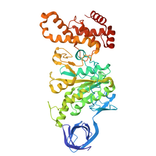
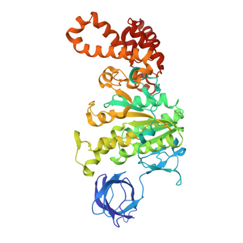
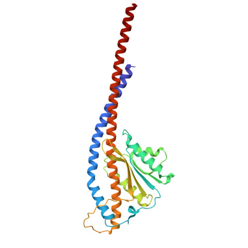
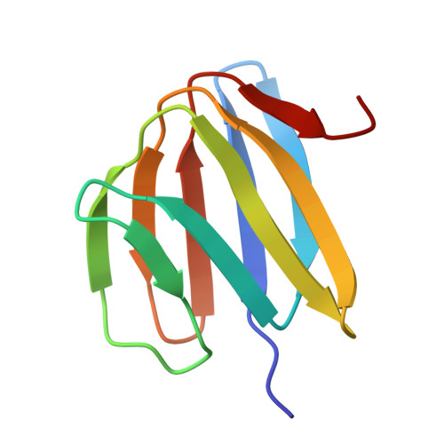
ADP-inhibited structure of non-catalytic site-depleted F o F 1 -ATPase from thermophilic Bacillus sp. PS-3.

Kobayashi, R.Nakano, A.Mitsuoka, K.Yokoyama, K.

(2025) Biochim Biophys Acta Bioenerg 1866: 149536-149536

  • DOI: https://doi.org/10.1016/j.bbabio.2025.149536
  • Primary Citation of Related Structures:  
    8YGV, 8YH8

  • PubMed Abstract: 

    The F 1 domain of F o F 1 -ATP synthases/ATPases (F o F 1 ) possesses three catalytic sites on the three αβ interfaces, termed α E β E , α D β D , and α T β T , located mainly on the β subunits. The enzyme also has three non-catalytic ATP-binding sites on the three αβ interfaces, located mainly on the α subunits. When ATP does not bind to the non-catalytic site, F o F 1 becomes significantly prone to ADP inhibition, ultimately resulting in the loss of ATPase activity. However, the underlying mechanism of ADP inhibition remains unclear. Here, we report the cryo-EM structure of the non-catalytic site-depleted (ΔNC) F o F 1 from thermophilic Bacillus sp. PS-3, which completely lacks the ability to bind ATP (and ADP) upon transitioning to the ADP-inhibited form. The structure closely resembled the 81° rotated structure of the wild-type F o F 1 , except for minor movements in the C-terminal region of the α subunit. In this structure, unlike the wild-type enzyme, the catalytic site at α D β D , responsible for ATP hydrolysis, was occupied by ADP-Mg, with the absence of Pi. Furthermore, the catalytic site at α E β E , where ATP enters the F 1 domain during steady-state catalysis, is occupied by ADP, seemingly impeding further ATP binding to the enzyme. The structure suggests that the ADP-inhibited form of the F 1 domain is more likely due to differences in the nucleotide-binding states at the catalytic sites rather than structural differences.


  • Organizational Affiliation
    • Department of Molecular Biosciences, Kyoto Sangyo University, Kamigamo-Motoyama, Kita-ku, Kyoto 603-8555, Japan.

Macromolecules
Find similar proteins by:  (by identity cutoff)  |  3D Structure
Entity ID: 1
MoleculeChains Sequence LengthOrganismDetailsImage
ATP synthase subunit alpha
A, B, C
476Bacillus sp. PS3Mutation(s): 0 
Gene Names: uncAatpA
EC: 7.1.2.2
UniProt
Find proteins for P09219 (Bacillus sp. (strain PS3))
Explore P09219 
Go to UniProtKB:  P09219
Entity Groups  
Sequence Clusters30% Identity50% Identity70% Identity90% Identity95% Identity100% Identity
UniProt GroupP09219
Sequence Annotations
Expand
  • Reference Sequence
Find similar proteins by:  (by identity cutoff)  |  3D Structure
Entity ID: 2
MoleculeChains Sequence LengthOrganismDetailsImage
ATP synthase subunit beta
D, E, F
471Bacillus sp. PS3Mutation(s): 0 
Gene Names: uncDatpD
EC: 7.1.2.2
UniProt
Find proteins for P07677 (Bacillus sp. (strain PS3))
Explore P07677 
Go to UniProtKB:  P07677
Entity Groups  
Sequence Clusters30% Identity50% Identity70% Identity90% Identity95% Identity100% Identity
UniProt GroupP07677
Sequence Annotations
Expand
  • Reference Sequence
Find similar proteins by:  (by identity cutoff)  |  3D Structure
Entity ID: 3
MoleculeChains Sequence LengthOrganismDetailsImage
ATP synthase gamma chain282Bacillus sp. PS3Mutation(s): 0 
Gene Names: uncGatpG
UniProt
Find proteins for P09222 (Bacillus sp. (strain PS3))
Explore P09222 
Go to UniProtKB:  P09222
Entity Groups  
Sequence Clusters30% Identity50% Identity70% Identity90% Identity95% Identity100% Identity
UniProt GroupP09222
Sequence Annotations
Expand
  • Reference Sequence
Find similar proteins by:  (by identity cutoff)  |  3D Structure
Entity ID: 4
MoleculeChains Sequence LengthOrganismDetailsImage
ATP synthase epsilon chain87Bacillus sp. PS3Mutation(s): 0 
Gene Names: uncCatpC
UniProt
Find proteins for P07678 (Bacillus sp. (strain PS3))
Explore P07678 
Go to UniProtKB:  P07678
Entity Groups  
Sequence Clusters30% Identity50% Identity70% Identity90% Identity95% Identity100% Identity
UniProt GroupP07678
Sequence Annotations
Expand
  • Reference Sequence
Small Molecules
Ligands 3 Unique
IDChains Name / Formula / InChI Key2D Diagram3D Interactions
ATP (Subject of Investigation/LOI)
Query on ATP

Download Ideal Coordinates CCD File 
M [auth F]ADENOSINE-5'-TRIPHOSPHATE
C10 H16 N5 O13 P3
ZKHQWZAMYRWXGA-KQYNXXCUSA-N
ADP (Subject of Investigation/LOI)
Query on ADP

Download Ideal Coordinates CCD File 
J [auth D],
K [auth E]
ADENOSINE-5'-DIPHOSPHATE
C10 H15 N5 O10 P2
XTWYTFMLZFPYCI-KQYNXXCUSA-N
MG (Subject of Investigation/LOI)
Query on MG

Download Ideal Coordinates CCD File 
I [auth D],
L [auth E],
N [auth F]
MAGNESIUM ION
Mg
JLVVSXFLKOJNIY-UHFFFAOYSA-N
Experimental Data & Validation

Experimental Data

  • Method: ELECTRON MICROSCOPY
  • Resolution: 2.70 Å
  • Aggregation State: PARTICLE 
  • Reconstruction Method: SINGLE PARTICLE 
EM Software:
TaskSoftware PackageVersion
RECONSTRUCTIONRELION4.0

Structure Validation

View Full Validation Report


Entry History & Funding Information

Deposition Data


Funding OrganizationLocationGrant Number
Other governmentJapan23H03231

Revision History  (Full details and data files)

  • Version 1.0: 2025-05-14
    Type: Initial release