Different DNA Binding and Damage Mode between Anticancer Antibiotics Trioxacarcin A and LL-D49194 alpha 1.
Gao, R.Q., Hu, X.D., Zhou, Q., Hou, X.F., Cao, C., Tang, G.L.(2024) JACS Au 4: 3641-3648
- PubMed: 39328742 Search on PubMedSearch on PubMed Central
- DOI: https://doi.org/10.1021/jacsau.4c00611
- Primary Citation Related Structures:
8XC7, 8XIC, 8Y1I - PubMed Abstract:
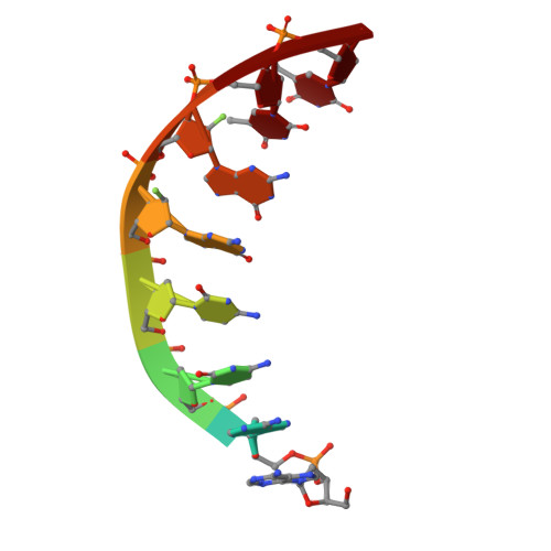
Trioxacarcin A (TXN) is a highly potent cytotoxic antibiotic with remarkable structural complexity. The crystal structure of TXN bound to double-stranded DNA (dsDNA) suggested that the TXN interaction might depend on positions of two sugar subunits on the minor and major grooves of dsDNA. LL-D49194α1 (LLD) is a TXN analogue bearing the same polycyclic polyketide scaffold with a distinct glycosylation pattern. Although LLD was in a phase I clinical trial, how LLD binds to dsDNA remains unclear. Here, we solved the solution structures at high resolutions of palindromic 2″-fluorine-labeled guanine-containing duplex d(A 1 A 2 C 3 C 4 G F G F T 7 T 8 ) 2 and of its stable LLD and TXN covalently bound complexes. Combined with biochemical assays, we found that TXN-alkylated dsDNA would tend to keep DNA helix conformation, while LLD-alkylated dsDNA lost its stability more than TXN-alkylated dsDNA, leading to dsDNA denaturation. Thus, despite lower cytotoxicity in vitro, the differences of sugar substitutions in LLD caused greater DNA damage than TXN, thereby bringing about a completely new biological effect.
- State Key Laboratory of Chemical Biology, Shanghai Institute of Organic Chemistry, University of Chinese Academy of Sciences, Chinese Academy of Sciences, Shanghai 200032, China.
Organizational Affiliation: