8Y0H | pdb_00008y0h

Structure of CXCR3 in complex with VUF11418 (Receptor-ligand focused map)


Experimental Data Snapshot

  • Method: ELECTRON MICROSCOPY
  • Resolution: 3.53 Å
  • Aggregation State: PARTICLE 
  • Reconstruction Method: SINGLE PARTICLE 

Starting Model: in silico
View more details

wwPDB Validation 3D Report Full Report

Validation slider image for 8Y0H

This is version 1.1 of the entry. See complete history

Literature

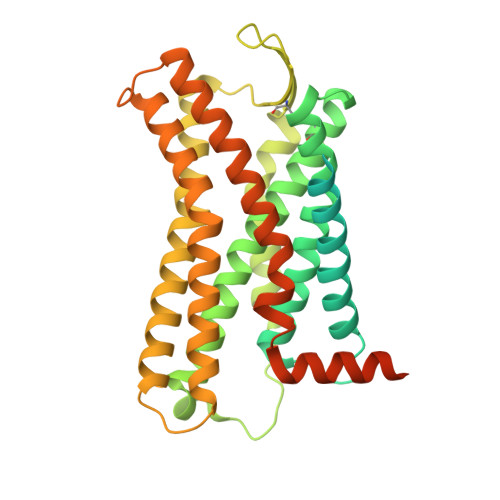
Structural visualization of small molecule recognition by CXCR3 uncovers dual-agonism in the CXCR3-CXCR7 system.

Saha, S.Sano, F.K.Sharma, S.Ganguly, M.Dalal, A.Mishra, S.Tiwari, D.Akasaka, H.Kobayashi, T.A.Roy, N.Zaidi, N.Itoh, Y.Leurs, R.Banerjee, R.Shihoya, W.Nureki, O.Shukla, A.K.

(2025) Nat Commun 16: 3047-3047

  • DOI: https://doi.org/10.1038/s41467-025-58264-w
  • Primary Citation Related Structures: 
    8XXY, 8XXZ, 8XYI, 8XYK, 8Y0H, 8Y0N

  • PubMed Abstract: 

    Chemokine receptors are critically involved in multiple physiological and pathophysiological processes related to immune response mechanisms. Most chemokine receptors are prototypical GPCRs although some also exhibit naturally-encoded signaling-bias toward β-arrestins (βarrs). C-X-C type chemokine receptors, namely CXCR3 and CXCR7, constitute a pair wherein the former is a prototypical GPCR while the latter exhibits selective coupling to βarrs despite sharing a common natural agonist: CXCL11. Moreover, CXCR3 and CXCR7 also recognize small molecule agonists suggesting a modular orthosteric ligand binding pocket. Here, we determine cryo-EM structures of CXCR3 in an Apo-state and in complex with small molecule agonists biased toward G-proteins or βarrs. These structural snapshots uncover an allosteric network bridging the ligand-binding pocket to intracellular side, driving the transducer-coupling bias at this receptor. Furthermore, structural topology of the orthosteric binding pocket also allows us to discover and validate that selected small molecule agonists of CXCR3 display robust agonism at CXCR7. Collectively, our study offers molecular insights into signaling-bias and dual agonism in the CXCR3-CXCR7 system with therapeutic implications.


  • Organizational Affiliation
    • Department of Biological Sciences and Bioengineering, Indian Institute of Technology Kanpur, Kanpur, India.

Macromolecule Content 

  • Total Structure Weight: 47.07 kDa 
  • Atom Count: 2,198 
  • Modeled Residue Count: 279 
  • Deposited Residue Count: 424 
  • Unique protein chains: 1

Macromolecules

Find similar proteins by:|  3D Structure
Entity ID: 1
MoleculeChains  Sequence LengthOrganismDetailsImage
C-X-C chemokine receptor type 3A [auth R]424Homo sapiensMutation(s): 0 
Gene Names: CXCR3
UniProt & NIH Common Fund Data Resources
Find proteins for P49682 (Homo sapiens)
Explore P49682 
Go to UniProtKB:  P49682
PHAROS:  P49682
GTEx:  ENSG00000186810 
Entity Groups
Sequence Clusters30% Identity50% Identity70% Identity90% Identity95% Identity100% Identity
UniProt GroupP49682
Sequence Annotations
Expand
Reference Sequence

Small Molecules

Ligands 1 Unique
IDChains Name / Formula / InChI Key2D Diagram3D Interactions
A1LW2
(Subject of Investigation/LOI)

Query on A1LW2


Download:Ideal Coordinates CCD File
B [auth R][(1~{R},5~{S})-6,6-dimethyl-2-bicyclo[3.1.1]hept-2-enyl]methyl-[[4-(2-iodanylphenyl)phenyl]methyl]-dimethyl-azanium
C25 H31 I N
FKUOVGQOKVAIAV-GMAHTHKFSA-N

Experimental Data & Validation

Experimental Data

  • Method: ELECTRON MICROSCOPY
  • Resolution: 3.53 Å
  • Aggregation State: PARTICLE 
  • Reconstruction Method: SINGLE PARTICLE 
EM Software:
TaskSoftware PackageVersion
RECONSTRUCTIONcryoSPARC4.4
MODEL REFINEMENTPHENIX

Structure Validation

View Full Validation Report


Entry History 

& Funding Information

Deposition Data


Funding OrganizationLocationGrant Number
Science and Engineering Research Board (SERB)IndiaIPA/2020/000405

Revision History  (Full details and data files)

  • Version 1.0: 2025-02-26
    Type: Initial release
  • Version 1.1: 2025-04-09
    Changes: Data collection, Database references