8XCJ

Open State of central tail fiber of bacteriophage lambda upon binding to LamB (gpJ713-LamB complex)


Experimental Data Snapshot

  • Method: ELECTRON MICROSCOPY
  • Resolution: 2.98 Å
  • Aggregation State: PARTICLE 
  • Reconstruction Method: SINGLE PARTICLE 

wwPDB Validation   3D Report Full Report


This is version 1.1 of the entry. See complete history


Literature

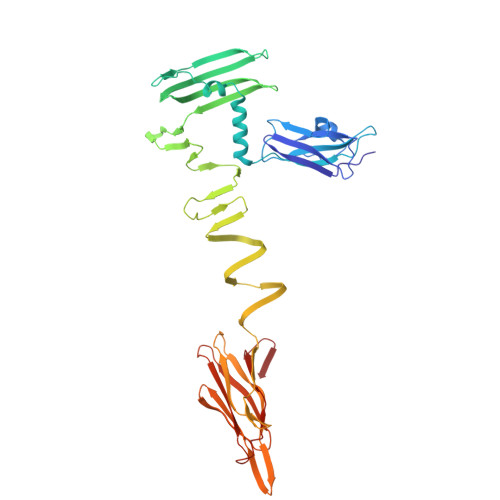
Structural mechanism of bacteriophage lambda tail's interaction with the bacterial receptor.

Ge, X.Wang, J.

(2024) Nat Commun 15: 4185-4185

  • DOI: https://doi.org/10.1038/s41467-024-48686-3
  • Primary Citation of Related Structures:  
    8XCG, 8XCI, 8XCJ, 8XCK

  • PubMed Abstract: 

    Bacteriophage infection, a pivotal process in microbiology, initiates with the phage's tail recognizing and binding to the bacterial cell surface, which then mediates the injection of viral DNA. Although comprehensive studies on the interaction between bacteriophage lambda and its outer membrane receptor, LamB, have provided rich information about the system's biochemical properties, the precise molecular mechanism remains undetermined. This study revealed the high-resolution cryo-electron microscopy (cryo-EM) structures of the bacteriophage lambda tail complexed with its irreversible Shigella sonnei 3070 LamB receptor and the closed central tail fiber. These structures reveal the complex processes that trigger infection and demonstrate a substantial conformational change in the phage lambda tail tip upon LamB binding. Providing detailed structures of bacteriophage lambda infection initiation, this study contributes to the expanding knowledge of lambda-bacterial interaction, which holds significance in the fields of microbiology and therapeutic development.


  • Organizational Affiliation

    State Key Laboratory of Membrane Biology, Beijing Frontier Research Center for Biological Structure, School of Life Sciences, Tsinghua University, 100084, Beijing, PR China.


Macromolecules
Find similar proteins by:  (by identity cutoff)  |  3D Structure
Entity ID: 1
MoleculeChains Sequence LengthOrganismDetailsImage
Maltoporin
A, B, C
422Shigella sonneiMutation(s): 0 
Gene Names: lamB
UniProt
Find proteins for A0A0I1R9L6 (Shigella sonnei)
Explore A0A0I1R9L6 
Go to UniProtKB:  A0A0I1R9L6
Entity Groups  
Sequence Clusters30% Identity50% Identity70% Identity90% Identity95% Identity100% Identity
UniProt GroupA0A0I1R9L6
Sequence Annotations
Expand
  • Reference Sequence
Find similar proteins by:  (by identity cutoff)  |  3D Structure
Entity ID: 2
MoleculeChains Sequence LengthOrganismDetailsImage
Tip attachment protein JD [auth Z],
E [auth J],
F
420Escherichia phage LambdaMutation(s): 0 
UniProt
Find proteins for P03749 (Escherichia phage lambda)
Explore P03749 
Go to UniProtKB:  P03749
Entity Groups  
Sequence Clusters30% Identity50% Identity70% Identity90% Identity95% Identity100% Identity
UniProt GroupP03749
Sequence Annotations
Expand
  • Reference Sequence
Experimental Data & Validation

Experimental Data

  • Method: ELECTRON MICROSCOPY
  • Resolution: 2.98 Å
  • Aggregation State: PARTICLE 
  • Reconstruction Method: SINGLE PARTICLE 

Structure Validation

View Full Validation Report


Entry History & Funding Information

Deposition Data


Funding OrganizationLocationGrant Number
National Natural Science Foundation of China (NSFC)China32371254
National Natural Science Foundation of China (NSFC)China32171190

Revision History  (Full details and data files)

  • Version 1.0: 2024-05-01
    Type: Initial release
  • Version 1.1: 2024-05-29
    Changes: Database references