8X5T | pdb_00008x5t

Crystal structure of Thermus thermophilus peptidyl-tRNA hydrolase in complex with adenosine 5'-monophosphate


Experimental Data Snapshot

  • Method: X-RAY DIFFRACTION
  • Resolution: 1.60 Å
  • R-Value Free: 
    0.168 (Depositor), 0.180 (DCC) 
  • R-Value Work: 
    0.144 (Depositor), 0.159 (DCC) 
  • R-Value Observed: 
    0.145 (Depositor) 

Starting Model: experimental
View more details

wwPDB Validation   3D Report Full Report


Ligand Structure Quality Assessment 


This is version 1.1 of the entry. See complete history


Literature

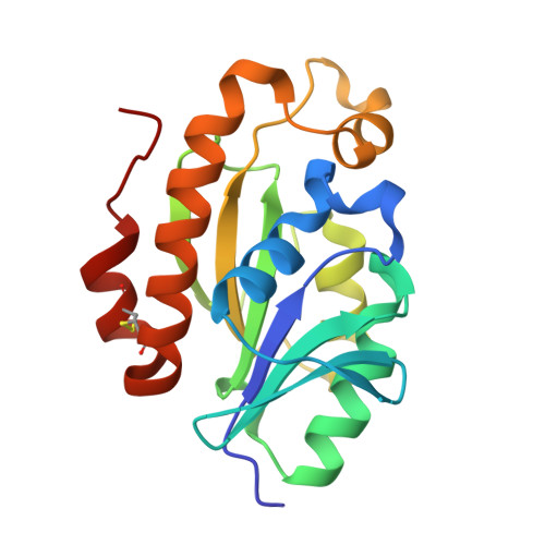
Binding mode between peptidyl-tRNA hydrolase and the peptidyl-A76 moiety of the substrate.

Uehara, Y.Matsumoto, A.Nakazawa, T.Fukuta, A.Ando, K.Uchiumi, T.Oka, N.Ito, K.

(2025) J Biological Chem 301: 108385-108385

  • DOI: https://doi.org/10.1016/j.jbc.2025.108385
  • Primary Citation of Related Structures:  
    8X5T, 8X5U

  • PubMed Abstract: 

    Peptidyl-tRNA hydrolase (Pth) hydrolyzes the ester bond between the peptide and the tRNA of peptidyl-tRNA molecules, which are the products of aborted translation, to prevent cell death by recycling tRNA. Numerous studies have attempted to elucidate the substrate recognition mechanism of Pth. However, the binding mode of the peptidyl-A76 (3'-terminal adenosine of tRNA) moiety of the substrate to Pth, especially the A76 moiety, remains unclear. Here, we present the crystal structure of Thermus thermophilus Pth (TtPth) in complex with adenosine 5'-monophosphate (AMP), a mimic of A76. In addition, we show the crystal structure of TtPth in which the active site cleft interacts with the C-terminal three amino acid residues of a crystallographically related neighboring TtPth molecule. Superimposition of these two crystal structures reveals that the C-terminal carboxyl group of the neighboring TtPth molecule and the 3'-hydroxyl group of AMP are located in positions favorable for ester bond formation, and we present a TtPth⋅peptidyl-A76 complex model. The complex model agrees with many previous NMR and kinetic studies, and our site-directed mutagenesis studies support its validity. Based on these facts, we conclude that the complex model properly represents the interaction between Pth and the substrate in the reaction. Furthermore, structural comparisons suggest that the substrate recognition mode is conserved among bacterial Pths. This study provides insights into the molecular mechanism of the reaction and useful information to design new drugs targeting Pth.


  • Organizational Affiliation
    • Department of Biology, Faculty of Science, Niigata University, Niigata, Japan.

Macromolecules
Find similar proteins by:  (by identity cutoff)  |  3D Structure
Entity ID: 1
MoleculeChains Sequence LengthOrganismDetailsImage
Peptidyl-tRNA hydrolase
A, B, C
187Thermus thermophilus HB8Mutation(s): 0 
Gene Names: pthTTHA1588
EC: 3.1.1.29
UniProt
Find proteins for Q5SHZ2 (Thermus thermophilus (strain ATCC 27634 / DSM 579 / HB8))
Explore Q5SHZ2 
Go to UniProtKB:  Q5SHZ2
Entity Groups  
Sequence Clusters30% Identity50% Identity70% Identity90% Identity95% Identity100% Identity
UniProt GroupQ5SHZ2
Sequence Annotations
Expand
  • Reference Sequence
Small Molecules
Ligands 3 Unique
IDChains Name / Formula / InChI Key2D Diagram3D Interactions
AMP (Subject of Investigation/LOI)
Query on AMP

Download Ideal Coordinates CCD File 
D [auth A],
H [auth B]
ADENOSINE MONOPHOSPHATE
C10 H14 N5 O7 P
UDMBCSSLTHHNCD-KQYNXXCUSA-N
TLA
Query on TLA

Download Ideal Coordinates CCD File 
E [auth A],
F [auth A]
L(+)-TARTARIC ACID
C4 H6 O6
FEWJPZIEWOKRBE-JCYAYHJZSA-N
GOL
Query on GOL

Download Ideal Coordinates CCD File 
G [auth A],
I [auth B],
J [auth B]
GLYCEROL
C3 H8 O3
PEDCQBHIVMGVHV-UHFFFAOYSA-N
Experimental Data & Validation

Experimental Data

  • Method: X-RAY DIFFRACTION
  • Resolution: 1.60 Å
  • R-Value Free:  0.168 (Depositor), 0.180 (DCC) 
  • R-Value Work:  0.144 (Depositor), 0.159 (DCC) 
  • R-Value Observed: 0.145 (Depositor) 
Space Group: P 61 2 2
Unit Cell:
Length ( Å )Angle ( ˚ )
a = 120.315α = 90
b = 120.315β = 90
c = 185.981γ = 120
Software Package:
Software NamePurpose
REFMACrefinement
HKL-2000data reduction
HKL-2000data scaling
MOLREPphasing

Structure Validation

View Full Validation Report


Ligand Structure Quality Assessment 


Entry History & Funding Information

Deposition Data


Funding OrganizationLocationGrant Number
Not funded--

Revision History  (Full details and data files)

  • Version 1.0: 2024-11-20
    Type: Initial release
  • Version 1.1: 2025-06-04
    Changes: Database references