8WKT

Cryo-EM structure of DSR2-DSAD1 complex


Experimental Data Snapshot

  • Method: ELECTRON MICROSCOPY
  • Resolution: 3.86 Å
  • Aggregation State: PARTICLE 
  • Reconstruction Method: SINGLE PARTICLE 

wwPDB Validation   3D Report Full Report


This is version 1.0 of the entry. See complete history


Literature

Molecular basis of bacterial DSR2 anti-phage defense and viral immune evasion.

Huang, J.Zhu, K.Gao, Y.Ye, F.Li, Z.Ge, Y.Liu, S.Yang, J.Gao, A.

(2024) Nat Commun 15: 3954-3954

  • DOI: https://doi.org/10.1038/s41467-024-48291-4
  • Primary Citation of Related Structures:  
    8WKS, 8WKT, 8WKX

  • PubMed Abstract: 

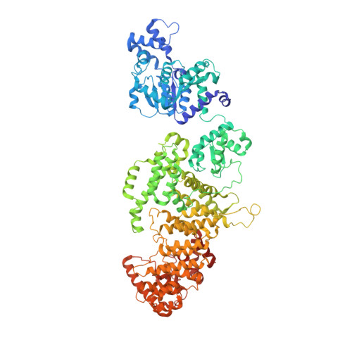
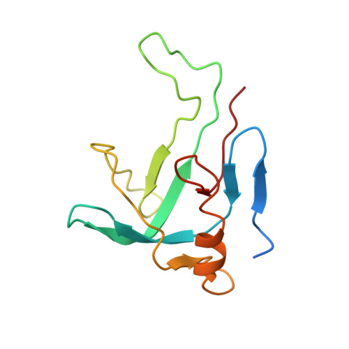
    Defense-associated sirtuin 2 (DSR2) systems are widely distributed across prokaryotic genomes, providing robust protection against phage infection. DSR2 recognizes phage tail tube proteins and induces abortive infection by depleting intracellular NAD + , a process that is counteracted by another phage-encoded protein, DSR Anti Defense 1 (DSAD1). Here, we present cryo-EM structures of Bacillus subtilis DSR2 in its apo, Tube-bound, and DSAD1-bound states. DSR2 assembles into an elongated tetramer, with four NADase catalytic modules clustered in the center and the regulatory-sensing modules distributed at four distal corners. Interestingly, monomeric Tube protein, rather than its oligomeric states, docks at each corner of the DSR2 tetramer to form a 4:4 DSR2-Tube assembly, which is essential for DSR2 NADase activity. DSAD1 competes with Tube for binding to DSR2 by occupying an overlapping region, thereby inhibiting DSR2 immunity. Thus, our results provide important insights into the assembly, activation and inhibition of the DSR2 anti-phage defense system.


  • Organizational Affiliation

    Key Laboratory of Molecular Medicine and Biotherapy, Aerospace Center Hospital, School of Life Science, Beijing Institute of Technology, Beijing, 100081, China. jfhuang@ibp.ac.cn.


Macromolecules
Find similar proteins by:  (by identity cutoff)  |  3D Structure
Entity ID: 1
MoleculeChains Sequence LengthOrganismDetailsImage
SIR2-like domain-containing protein
A, B, D, E
1,004Bacillus subtilis subsp. natto BEST195Mutation(s): 0 
Gene Names: BSNT_07056
UniProt
Find proteins for D4G637 (Bacillus subtilis subsp. natto (strain BEST195))
Explore D4G637 
Go to UniProtKB:  D4G637
Entity Groups  
Sequence Clusters30% Identity50% Identity70% Identity90% Identity95% Identity100% Identity
UniProt GroupD4G637
Sequence Annotations
Expand
  • Reference Sequence
Find similar proteins by:  (by identity cutoff)  |  3D Structure
Entity ID: 2
MoleculeChains Sequence LengthOrganismDetailsImage
SPbeta prophage-derived uncharacterized protein YotI120CaudoviricetesMutation(s): 0 
Gene Names: yotIyodWyokFBSU19870
UniProt
Find proteins for Q796A8 (Bacillus subtilis (strain 168))
Explore Q796A8 
Go to UniProtKB:  Q796A8
Entity Groups  
Sequence Clusters30% Identity50% Identity70% Identity90% Identity95% Identity100% Identity
UniProt GroupQ796A8
Sequence Annotations
Expand
  • Reference Sequence
Experimental Data & Validation

Experimental Data

  • Method: ELECTRON MICROSCOPY
  • Resolution: 3.86 Å
  • Aggregation State: PARTICLE 
  • Reconstruction Method: SINGLE PARTICLE 
EM Software:
TaskSoftware PackageVersion
MODEL REFINEMENTPHENIX1.20.1_4487:

Structure Validation

View Full Validation Report


Entry History & Funding Information

Deposition Data


Funding OrganizationLocationGrant Number
Not funded--

Revision History  (Full details and data files)

  • Version 1.0: 2024-05-22
    Type: Initial release