


Structural Flexibility of the Monomeric Red Fluorescent Protein DsRed.
Nam, K.H.(2024) Crystals (Basel) 14
Experimental Data Snapshot
Starting Model: experimental
View more details
wwPDB Validation 3D Report Full Report

(2024) Crystals (Basel) 14
Entity ID: 1 | |||||
|---|---|---|---|---|---|
| Molecule | Chains | Sequence Length | Organism | Details | Image |
| Red fluorescent protein | 223 | Discosoma sp. | Mutation(s): 0 | 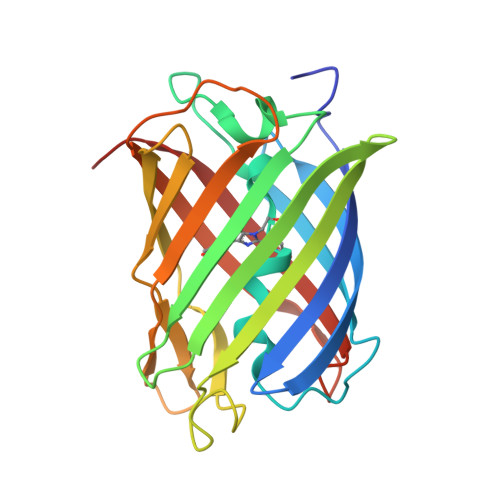 | |
| Modified Residues 1 Unique | |||||
|---|---|---|---|---|---|
| ID | Chains | Type | Formula | 2D Diagram | Parent |
| CRQ Query on CRQ | A, B | L-PEPTIDE LINKING | C16 H16 N4 O5 |  | GLN, TYR, GLY |
| Length ( Å ) | Angle ( ˚ ) |
|---|---|
| a = 39.566 | α = 90 |
| b = 107.001 | β = 104.87 |
| c = 50.027 | γ = 90 |
| Software Name | Purpose |
|---|---|
| REFMAC | refinement |
| HKL-2000 | data reduction |
| HKL-2000 | data scaling |
| MOLREP | phasing |
| Funding Organization | Location | Grant Number |
|---|---|---|
| National Research Foundation (NRF, Korea) | Korea, Republic Of | NRF-2021R1I1A1A01050838 |


