8VDQ | pdb_00008vdq

Cryogenic electron microscopy model of full-length talin


Experimental Data Snapshot

  • Method: ELECTRON MICROSCOPY
  • Resolution: 5.50 Å
  • Aggregation State: PARTICLE 
  • Reconstruction Method: SINGLE PARTICLE 

Starting Model: experimental
View more details

wwPDB Validation   3D Report Full Report


This is version 1.1 of the entry. See complete history


Literature

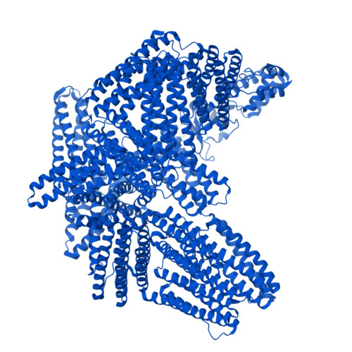
High-resolution snapshots of the talin auto-inhibitory states suggest roles in cell adhesion and signaling.

Rangarajan, E.S.Bois, J.L.Hansen, S.B.Izard, T.

(2024) Nat Commun 15: 9270-9270

  • DOI: https://doi.org/10.1038/s41467-024-52581-2
  • Primary Citation of Related Structures:  
    8VDO, 8VDP, 8VDQ, 8VDR

  • PubMed Abstract: 

    Talin regulates crucial cellular functions, including cell adhesion and motility, and affects human diseases. Triggered by mechanical forces, talin plays crucial roles in facilitating the formation of focal adhesions and recruiting essential focal adhesion regulatory elements such as vinculin. The structural flexibility allows talin to fine-tune its signaling responses. This study presents our 2.7 Å cryoEM structures of talin, which surprisingly uncovers several auto-inhibitory states. Contrary to previous suggestions, our structures reveal that (1) the first and last three domains are not involved in maintaining talin in its closed state and are mobile, (2) the talin F-actin and membrane binding domain are loosely attached and thus available for binding, and (3) the main force-sensing domain is oriented with its vinculin binding sites ready for release. These structural snapshots offer insights and advancements in understanding the dynamic talin activation mechanism, which is crucial for mediating cell adhesion.


  • Organizational Affiliation
    • Cell Adhesion Laboratory, UF Scripps, Jupiter, FL, USA.

Macromolecules
Find similar proteins by:  (by identity cutoff)  |  3D Structure
Entity ID: 1
MoleculeChains Sequence LengthOrganismDetailsImage
Green fluorescent protein,Talin-12,804Mus musculusMutation(s): 0 
Gene Names: GFPTln1Tln
UniProt
Find proteins for P26039 (Mus musculus)
Explore P26039 
Go to UniProtKB:  P26039
Find proteins for P42212 (Aequorea victoria)
Explore P42212 
Go to UniProtKB:  P42212
Entity Groups  
Sequence Clusters30% Identity50% Identity70% Identity90% Identity95% Identity100% Identity
UniProt GroupsP42212P26039
Sequence Annotations
Expand
  • Reference Sequence
Experimental Data & Validation

Experimental Data

  • Method: ELECTRON MICROSCOPY
  • Resolution: 5.50 Å
  • Aggregation State: PARTICLE 
  • Reconstruction Method: SINGLE PARTICLE 
EM Software:
TaskSoftware PackageVersion
MODEL REFINEMENTPHENIX1.18.2_3874:
RECONSTRUCTIONcryoSPARC

Structure Validation

View Full Validation Report


Entry History & Funding Information

Deposition Data


Funding OrganizationLocationGrant Number
National Science Foundation (NSF, United States)United States--

Revision History  (Full details and data files)

  • Version 1.0: 2024-10-02
    Type: Initial release
  • Version 1.1: 2025-08-06
    Changes: Data collection, Database references, Structure summary