8VDA

Crystal structure of Bacillus subtilis FabHA-coenzyme A complex

  • Classification: TRANSFERASE
  • Organism(s): Bacillus subtilis
  • Expression System: Escherichia coli
  • Mutation(s): No 

  • Deposited: 2023-12-14 Released: 2024-02-14 
  • Deposition Author(s): Radka, C.D., Rock, C.O.
  • Funding Organization(s): National Institutes of Health/National Institute Of Allergy and Infectious Diseases (NIH/NIAID), National Institutes of Health/National Institute of General Medical Sciences (NIH/NIGMS)

Experimental Data Snapshot

  • Method: X-RAY DIFFRACTION
  • Resolution: 2.02 Å
  • R-Value Free: 0.231 
  • R-Value Work: 0.187 
  • R-Value Observed: 0.192 

wwPDB Validation   3D Report Full Report


Ligand Structure Quality Assessment 


This is version 1.0 of the entry. See complete history


Literature

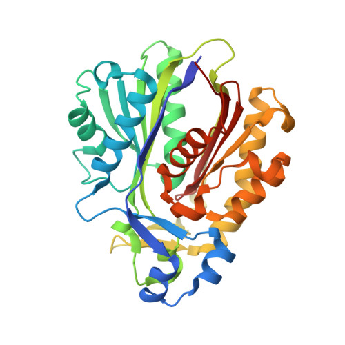
Crystal structures of the fatty acid biosynthesis initiation enzymes in Bacillus subtilis.

Radka, C.D.Rock, C.O.

(2024) J Struct Biol 216: 108065-108065

  • DOI: https://doi.org/10.1016/j.jsb.2024.108065
  • Primary Citation of Related Structures:  
    8VD9, 8VDA, 8VDB

  • PubMed Abstract: 

    Bacteria use the fatty acid composition of membrane lipids to maintain homeostasis of the bilayer. β-Ketoacyl-ACP synthase III (FabH) initiates fatty acid biosynthesis and is the primary determinant of the fatty acid composition. FabH condenses malonyl-acyl carrier protein with an acyl-Coenzyme A primer to form β -ketoacyl-acyl carrier protein which is used to make substrates for lipid synthesis. The acyl-Coenzyme A primer determines whether an acyl chain in the membrane has iso, anteiso, or no branching (straight chain) and biophysical properties of the membrane. The soil bacterium Bacillus subtilis encodes two copies of FabH (BsFabHA and BsFabHB), and here we solve their crystal structures. The substrate-free 1.85 Å and 2.40 Å structures of BsFabHA and BsFabHB show both enzymes have similar residues that line the active site but differ in the architecture surrounding the catalytic residues and oxyanion hole. Branching in the BsFabHB active site may better accommodate the structure of an iso-branched acyl-Coenzyme A molecule and thus confer superior utilization to BsFabHA for this primer type. The 2.02 Å structure of BsFabHA•Coenzyme A shows how the active site architecture changes after binding the first substrate. The other notable difference is an amino acid insertion in BsFabHB that extends a cap that covers the dimer interface. The cap topology is diverse across FabH structures and appears to be a distinguishing feature. FabH enzymes have variable sensitivity to natural product inhibitors and the availability of crystal structures help clarify how nature designs antimicrobials that differentially target FabH homologs.


  • Organizational Affiliation

    Department of Microbiology, Immunology, and Molecular Genetics, University of Kentucky, 760 Press Avenue, Lexington, KY 40536, USA. Electronic address: Christopher.radka@uky.edu.


Macromolecules
Find similar proteins by:  (by identity cutoff)  |  3D Structure
Entity ID: 1
MoleculeChains Sequence LengthOrganismDetailsImage
Beta-ketoacyl-[acyl-carrier-protein] synthase III 1317Bacillus subtilisMutation(s): 0 
Gene Names: fabHAfabHfabH1yjaXBSU11330
EC: 2.3.1.180 (PDB Primary Data), 2.3.1.300 (PDB Primary Data)
UniProt
Find proteins for O34746 (Bacillus subtilis (strain 168))
Explore O34746 
Go to UniProtKB:  O34746
Entity Groups  
Sequence Clusters30% Identity50% Identity70% Identity90% Identity95% Identity100% Identity
UniProt GroupO34746
Sequence Annotations
Expand
  • Reference Sequence
Small Molecules
Ligands 1 Unique
IDChains Name / Formula / InChI Key2D Diagram3D Interactions
COA (Subject of Investigation/LOI)
Query on COA

Download Ideal Coordinates CCD File 
B [auth A]COENZYME A
C21 H36 N7 O16 P3 S
RGJOEKWQDUBAIZ-IBOSZNHHSA-N
Experimental Data & Validation

Experimental Data

  • Method: X-RAY DIFFRACTION
  • Resolution: 2.02 Å
  • R-Value Free: 0.231 
  • R-Value Work: 0.187 
  • R-Value Observed: 0.192 
  • Space Group: P 41 21 2
Unit Cell:
Length ( Å )Angle ( ˚ )
a = 60.61α = 90
b = 60.61β = 90
c = 153.4γ = 90
Software Package:
Software NamePurpose
PHENIXrefinement
XDSdata reduction
XDSdata scaling
PHASERphasing

Structure Validation

View Full Validation Report


Ligand Structure Quality Assessment 


Entry History & Funding Information

Deposition Data


Funding OrganizationLocationGrant Number
National Institutes of Health/National Institute Of Allergy and Infectious Diseases (NIH/NIAID)United StatesAI166116
National Institutes of Health/National Institute of General Medical Sciences (NIH/NIGMS)United StatesGM034496

Revision History  (Full details and data files)

  • Version 1.0: 2024-02-14
    Type: Initial release