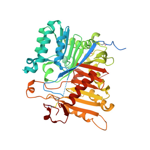
8V07

Crystal structure of mouse PLD3 co-crystallized with 5'Pi-ssDNA for 30 days

  • Classification: HYDROLASE
  • Organism(s): Mus musculus
  • Expression System: Homo sapiens
  • Mutation(s): No 

  • Deposited: 2023-11-17 Released: 2024-03-13 
  • Deposition Author(s): Yuan, M., Wilson, I.A.
  • Funding Organization(s): National Institutes of Health/National Institute Of Allergy and Infectious Diseases (NIH/NIAID), National Institutes of Health/National Institute on Aging (NIH/NIA)

Experimental Data Snapshot

  • Method: X-RAY DIFFRACTION
  • Resolution: 1.99 Å
  • R-Value Free: 0.264 
  • R-Value Work: 0.224 
  • R-Value Observed: 0.226 

wwPDB Validation   3D Report Full Report


Ligand Structure Quality Assessment 


This is version 1.1 of the entry. See complete history


Literature

Structural and mechanistic insights into disease-associated endolysosomal exonucleases PLD3 and PLD4.

Yuan, M.Peng, L.Huang, D.Gavin, A.Luan, F.Tran, J.Feng, Z.Zhu, X.Matteson, J.Wilson, I.A.Nemazee, D.

(2024) Structure 

  • DOI: https://doi.org/10.1016/j.str.2024.02.019
  • Primary Citation of Related Structures:  
    8V05, 8V06, 8V07, 8V08

  • PubMed Abstract: 

    Endolysosomal exonucleases PLD3 and PLD4 (phospholipases D3 and D4) are associated with autoinflammatory and autoimmune diseases. We report structures of these enzymes, and the molecular basis of their catalysis. The structures reveal an intra-chain dimer topology forming a basic active site at the interface. Like other PLD superfamily members, PLD3 and PLD4 carry HxKxxxxD/E motifs and participate in phosphodiester-bond cleavage. The enzymes digest ssDNA and ssRNA in a 5'-to-3' manner and are blocked by 5'-phosphorylation. We captured structures in apo, intermediate, and product states and revealed a "link-and-release" two-step catalysis. We also unexpectedly demonstrated phosphatase activity via a covalent 3-phosphohistidine intermediate. PLD4 contains an extra hydrophobic clamp that stabilizes substrate and could affect oligonucleotide substrate preference and product release. Biochemical and structural analysis of disease-associated mutants of PLD3/4 demonstrated reduced enzyme activity or thermostability and the possible basis for disease association. Furthermore, these findings provide insight into therapeutic design.


  • Organizational Affiliation

    Department of Integrative Structural and Computational Biology, The Scripps Research Institute, La Jolla, CA 92037, USA. Electronic address: myuan@scripps.edu.


Macromolecules
Find similar proteins by:  (by identity cutoff)  |  3D Structure
Entity ID: 1
MoleculeChains Sequence LengthOrganismDetailsImage
5'-3' exonuclease PLD3A [auth B],
B [auth A],
C,
D
467Mus musculusMutation(s): 0 
Gene Names: Pld3
UniProt & NIH Common Fund Data Resources
Find proteins for O35405 (Mus musculus)
Explore O35405 
Go to UniProtKB:  O35405
IMPC:  MGI:1333782
Entity Groups  
Sequence Clusters30% Identity50% Identity70% Identity90% Identity95% Identity100% Identity
UniProt GroupO35405
Sequence Annotations
Expand
  • Reference Sequence
Oligosaccharides

Help

Entity ID: 2
MoleculeChains Length2D Diagram Glycosylation3D Interactions
alpha-D-mannopyranose-(1-3)-[alpha-D-mannopyranose-(1-6)]alpha-D-mannopyranose-(1-6)-[alpha-D-mannopyranose-(1-3)]beta-D-mannopyranose-(1-4)-2-acetamido-2-deoxy-beta-D-glucopyranose-(1-4)-2-acetamido-2-deoxy-beta-D-glucopyranose
E, H
7N-Glycosylation
Glycosylation Resources
GlyTouCan:  G55220VL
GlyCosmos:  G55220VL
GlyGen:  G55220VL
Entity ID: 3
MoleculeChains Length2D Diagram Glycosylation3D Interactions
2-acetamido-2-deoxy-beta-D-glucopyranose-(1-4)-2-acetamido-2-deoxy-beta-D-glucopyranose
F, G, J, K
2N-Glycosylation
Glycosylation Resources
GlyTouCan:  G42666HT
GlyCosmos:  G42666HT
GlyGen:  G42666HT
Entity ID: 4
MoleculeChains Length2D Diagram Glycosylation3D Interactions
alpha-D-mannopyranose-(1-6)-beta-D-mannopyranose-(1-4)-2-acetamido-2-deoxy-beta-D-glucopyranose-(1-4)-2-acetamido-2-deoxy-beta-D-glucopyranose
I
4N-Glycosylation
Glycosylation Resources
GlyTouCan:  G22573RC
GlyCosmos:  G22573RC
GlyGen:  G22573RC
Small Molecules
Ligands 5 Unique
IDChains Name / Formula / InChI Key2D Diagram3D Interactions
NAG
Query on NAG

Download Ideal Coordinates CCD File 
Q [auth A]2-acetamido-2-deoxy-beta-D-glucopyranose
C8 H15 N O6
OVRNDRQMDRJTHS-FMDGEEDCSA-N
CIT
Query on CIT

Download Ideal Coordinates CCD File 
L [auth B]CITRIC ACID
C6 H8 O7
KRKNYBCHXYNGOX-UHFFFAOYSA-N
PO4
Query on PO4

Download Ideal Coordinates CCD File 
P [auth B],
S [auth A],
U [auth C],
X [auth D]
PHOSPHATE ION
O4 P
NBIIXXVUZAFLBC-UHFFFAOYSA-K
GOL
Query on GOL

Download Ideal Coordinates CCD File 
M [auth B]
O [auth B]
R [auth A]
T [auth C]
V [auth D]
M [auth B],
O [auth B],
R [auth A],
T [auth C],
V [auth D],
W [auth D]
GLYCEROL
C3 H8 O3
PEDCQBHIVMGVHV-UHFFFAOYSA-N
ACE
Query on ACE

Download Ideal Coordinates CCD File 
N [auth B]ACETYL GROUP
C2 H4 O
IKHGUXGNUITLKF-UHFFFAOYSA-N
Experimental Data & Validation

Experimental Data

  • Method: X-RAY DIFFRACTION
  • Resolution: 1.99 Å
  • R-Value Free: 0.264 
  • R-Value Work: 0.224 
  • R-Value Observed: 0.226 
  • Space Group: P 1
Unit Cell:
Length ( Å )Angle ( ˚ )
a = 54.523α = 83.65
b = 54.789β = 89.1
c = 203.574γ = 89.9
Software Package:
Software NamePurpose
PHENIXrefinement
HKL-2000data reduction
HKL-2000data scaling
PHASERphasing

Structure Validation

View Full Validation Report


Ligand Structure Quality Assessment 


Entry History & Funding Information

Deposition Data


Funding OrganizationLocationGrant Number
National Institutes of Health/National Institute Of Allergy and Infectious Diseases (NIH/NIAID)United StatesAI142945
National Institutes of Health/National Institute on Aging (NIH/NIA)United StatesAG070775

Revision History  (Full details and data files)

  • Version 1.0: 2024-03-13
    Type: Initial release
  • Version 1.1: 2024-04-17
    Changes: Database references