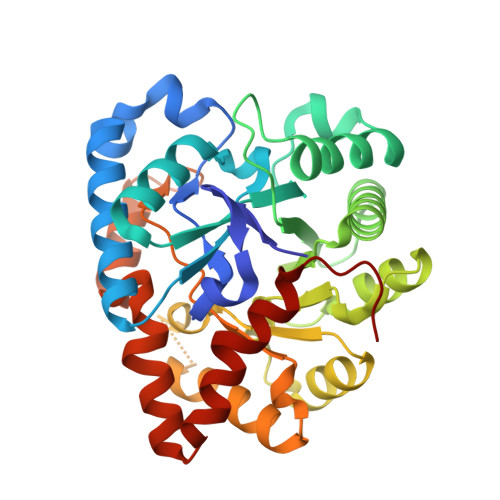
8UQZ

Round 18 Arylesterase Variant of Phosphotriesterase Bound to Gadolinium(III) Measured at 9.5 keV


Experimental Data Snapshot

  • Method: X-RAY DIFFRACTION
  • Resolution: 1.61 Å
  • R-Value Free: 0.195 
  • R-Value Work: 0.170 
  • R-Value Observed: 0.172 

wwPDB Validation   3D Report Full Report


This is version 1.1 of the entry. See complete history


Literature

Mononuclear binding and catalytic activity of europium(III) and gadolinium(III) at the active site of the model metalloenzyme phosphotriesterase.

Breeze, C.W.Nakano, Y.Campbell, E.C.Frkic, R.L.Lupton, D.W.Jackson, C.J.

(2024) Acta Crystallogr D Struct Biol 80: 289-298

  • DOI: https://doi.org/10.1107/S2059798324002316
  • Primary Citation of Related Structures:  
    8UQW, 8UQX, 8UQY, 8UQZ

  • PubMed Abstract: 

    Lanthanide ions have ideal chemical properties for catalysis, such as hard Lewis acidity, fast ligand-exchange kinetics, high coordination-number preferences and low geometric requirements for coordination. As a result, many small-molecule lanthanide catalysts have been described in the literature. Yet, despite the ability of enzymes to catalyse highly stereoselective reactions under gentle conditions, very few lanthanoenzymes have been investigated. In this work, the mononuclear binding of europium(III) and gadolinium(III) to the active site of a mutant of the model enzyme phosphotriesterase are described using X-ray crystallography at 1.78 and 1.61 Å resolution, respectively. It is also shown that despite coordinating a single non-natural metal cation, the PTE-R18 mutant is still able to maintain esterase activity.


  • Organizational Affiliation

    Research School of Chemistry, Australian National University, Canberra, ACT 2601, Australia.


Macromolecules
Find similar proteins by:  (by identity cutoff)  |  3D Structure
Entity ID: 1
MoleculeChains Sequence LengthOrganismDetailsImage
Phosphotriesterase variant PTE-R18
A, B
333Brevundimonas diminutaMutation(s): 0 
UniProt
Find proteins for A0A060GYS7 (Brevundimonas diminuta)
Explore A0A060GYS7 
Go to UniProtKB:  A0A060GYS7
Entity Groups  
Sequence Clusters30% Identity50% Identity70% Identity90% Identity95% Identity100% Identity
UniProt GroupA0A060GYS7
Sequence Annotations
Expand
  • Reference Sequence
Experimental Data & Validation

Experimental Data

  • Method: X-RAY DIFFRACTION
  • Resolution: 1.61 Å
  • R-Value Free: 0.195 
  • R-Value Work: 0.170 
  • R-Value Observed: 0.172 
  • Space Group: P 21 21 2
Unit Cell:
Length ( Å )Angle ( ˚ )
a = 85.714α = 90
b = 86.138β = 90
c = 89.247γ = 90
Software Package:
Software NamePurpose
PHENIXrefinement
Aimlessdata scaling
PHASERphasing
XDSdata reduction

Structure Validation

View Full Validation Report


Entry History & Funding Information

Deposition Data


Funding OrganizationLocationGrant Number
Australian Research Council (ARC)AustraliaCE200100012

Revision History  (Full details and data files)

  • Version 1.0: 2024-04-03
    Type: Initial release
  • Version 1.1: 2024-04-10
    Changes: Database references