Directing polymorph specific calcium carbonate formation with de novo protein templates.
Davila-Hernandez, F.A., Jin, B., Pyles, H., Zhang, S., Wang, Z., Huddy, T.F., Bera, A.K., Kang, A., Chen, C.L., De Yoreo, J.J., Baker, D.(2023) Nat Commun 14: 8191-8191
- PubMed: 38097544 Search on PubMedSearch on PubMed Central
- DOI: https://doi.org/10.1038/s41467-023-43608-1
- Primary Citation Related Structures:
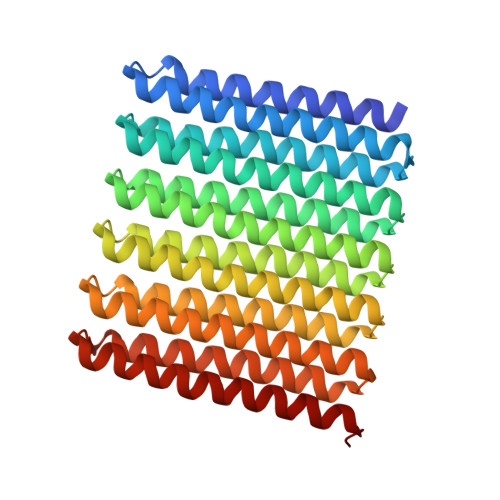
8UGC - PubMed Abstract:
Biomolecules modulate inorganic crystallization to generate hierarchically structured biominerals, but the atomic structure of the organic-inorganic interfaces that regulate mineralization remain largely unknown. We hypothesized that heterogeneous nucleation of calcium carbonate could be achieved by a structured flat molecular template that pre-organizes calcium ions on its surface. To test this hypothesis, we design helical repeat proteins (DHRs) displaying regularly spaced carboxylate arrays on their surfaces and find that both protein monomers and protein-Ca 2+ supramolecular assemblies directly nucleate nano-calcite with non-natural {110} or {202} faces while vaterite, which forms first in the absence of the proteins, is bypassed. These protein-stabilized nanocrystals then assemble by oriented attachment into calcite mesocrystals. We find further that nanocrystal size and polymorph can be tuned by varying the length and surface chemistry of the designed protein templates. Thus, bio-mineralization can be programmed using de novo protein design, providing a route to next-generation hybrid materials.
- Department of Biochemistry, University of Washington, Seattle, WA, 98195, USA.
Organizational Affiliation: