Capturing excited-state structural snapshots of evolutionary green-to-red photochromic fluorescent proteins.
Krueger, T.D., Henderson, J.N., Breen, I.L., Zhu, L., Wachter, R.M., Mills, J.H., Fang, C.(2023) Front Chem 11: 1328081-1328081
- PubMed: 38144887 Search on PubMedSearch on PubMed Central
- DOI: https://doi.org/10.3389/fchem.2023.1328081
- Primary Citation Related Structures:
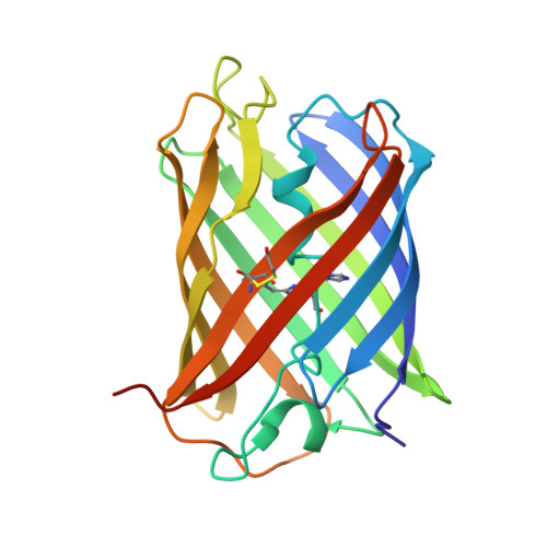
8THS, 8UB6 - PubMed Abstract:
Photochromic fluorescent proteins (FPs) have proved to be indispensable luminous probes for sophisticated and advanced bioimaging techniques. Among them, an interplay between photoswitching and photoconversion has only been observed in a limited subset of Kaede-like FPs that show potential for discovering the key mechanistic steps during green-to-red photoconversion. Various spectroscopic techniques including femtosecond stimulated Raman spectroscopy (FSRS), X-ray crystallography, and femtosecond transient absorption were employed on a set of five related FPs with varying photoconversion and photoswitching efficiencies. A 3-methyl-histidine chromophore derivative, incorporated through amber suppression using orthogonal aminoacyl tRNA synthetase/tRNA pairs, displays more dynamic photoswitching but greatly reduced photoconversion versus the least-evolved ancestor (LEA). Excitation-dependent measurements of the green anionic chromophore reveal that the varying photoswitching efficiencies arise from both the initial transient dynamics of the bright cis state and the final trans -like photoswitched off state, with an exocyclic bridge H-rocking motion playing an active role during the excited-state energy dissipation. This investigation establishes a close-knit feedback loop between spectroscopic characterization and protein engineering, which may be especially beneficial to develop more versatile FPs with targeted mutations and enhanced functionalities, such as photoconvertible FPs that also feature photoswitching properties.
- Department of Chemistry, Oregon State University, Corvallis, OR, United States.
Organizational Affiliation: