8U0P

Synaptic complex of human DNA polymerase Lambda DL variant engaged on a noncomplementary DNA double-strand break


Experimental Data Snapshot

  • Method: X-RAY DIFFRACTION
  • Resolution: 1.90 Å
  • R-Value Free: 0.193 
  • R-Value Work: 0.172 
  • R-Value Observed: 0.173 

wwPDB Validation   3D Report Full Report


Ligand Structure Quality Assessment 


This is version 1.0 of the entry. See complete history


Literature

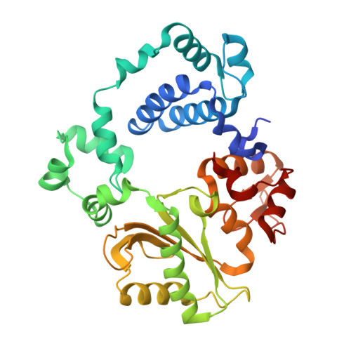
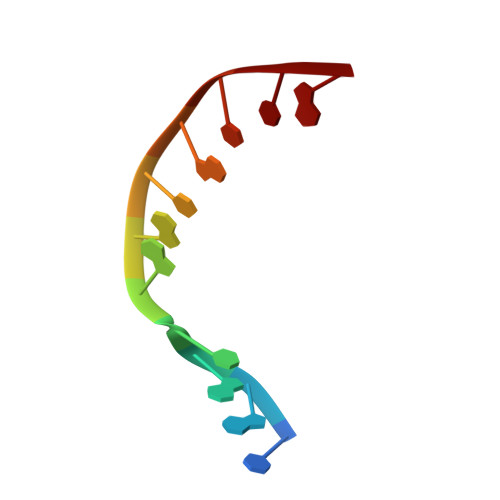
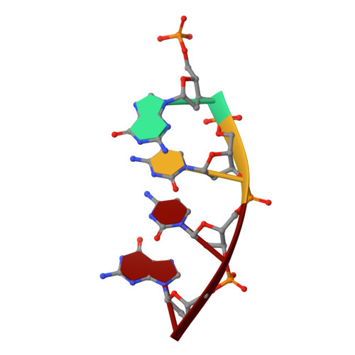
DNA polymerase lambda Loop1 variant yields unexpected gain-of-function capabilities in nonhomologous end-joining.

Kaminski, A.M.Chiruvella, K.K.Ramsden, D.A.Bebenek, K.Kunkel, T.A.Pedersen, L.C.

(2024) DNA Repair (Amst) 136: 103645-103645

  • DOI: https://doi.org/10.1016/j.dnarep.2024.103645
  • Primary Citation of Related Structures:  
    8U0O, 8U0P

  • PubMed Abstract: 

    DNA polymerases lambda (Polλ) and mu (Polμ) are X-Family polymerases that participate in DNA double-strand break (DSB) repair by the nonhomologous end-joining pathway (NHEJ). Both polymerases direct synthesis from one DSB end, using template derived from a second DSB end. In this way, they promote the NHEJ ligation step and minimize the sequence loss normally associated with this pathway. The two polymerases differ in cognate substrate, as Polλ is preferred when synthesis must be primed from a base-paired DSB end, while Polμ is required when synthesis must be primed from an unpaired DSB end. We generated a Polλ variant (Polλ KGET ) that retained canonical Polλ activity on a paired end-albeit with reduced incorporation fidelity. We recently discovered that the variant had unexpectedly acquired the activity previously unique to Polμ-synthesis from an unpaired primer terminus. Though the sidechains of the Loop1 region make no contact with the DNA substrate, Polλ KGET Loop1 amino acid sequence is surprisingly essential for its unique activity during NHEJ. Taken together, these results underscore that the Loop1 region plays distinct roles in different Family X polymerases.


  • Organizational Affiliation

    Genome Integrity and Structural Biology Laboratory, National Institute of Environmental Health Sciences, National Institutes of Health, 111 TW Alexander Dr., Bldg 101, Research Triangle Park, NC 27709, USA.


Macromolecules

Find similar proteins by:  (by identity cutoff)  |  3D Structure
Entity ID: 1
MoleculeChains Sequence LengthOrganismDetailsImage
DNA polymerase lambda340Homo sapiensMutation(s): 0 
Gene Names: POLL
EC: 2.7.7.7 (PDB Primary Data), 4.2.99 (PDB Primary Data)
UniProt & NIH Common Fund Data Resources
Find proteins for Q9UGP5 (Homo sapiens)
Explore Q9UGP5 
Go to UniProtKB:  Q9UGP5
PHAROS:  Q9UGP5
GTEx:  ENSG00000166169 
Entity Groups  
Sequence Clusters30% Identity50% Identity70% Identity90% Identity95% Identity100% Identity
UniProt GroupQ9UGP5
Sequence Annotations
Expand
  • Reference Sequence

Find similar nucleic acids by:  Sequence   |   3D Structure  

Entity ID: 2
MoleculeChains LengthOrganismImage
DNA (5'-D(*CP*GP*GP*CP*AP*GP*TP*AP*CP*TP*G)-3')B [auth T]11synthetic construct
Sequence Annotations
Expand
  • Reference Sequence

Find similar nucleic acids by:  Sequence   |   3D Structure  

Entity ID: 3
MoleculeChains LengthOrganismImage
DNA (5'-D(*CP*AP*GP*TP*AP*C)-3')C [auth P]6synthetic construct
Sequence Annotations
Expand
  • Reference Sequence

Find similar nucleic acids by:  Sequence   |   3D Structure  

Entity ID: 4
MoleculeChains LengthOrganismImage
DNA (5'-D(P*GP*CP*CP*G)-3')4synthetic construct
Sequence Annotations
Expand
  • Reference Sequence
Small Molecules
Ligands 5 Unique
IDChains Name / Formula / InChI Key2D Diagram3D Interactions
DUP (Subject of Investigation/LOI)
Query on DUP

Download Ideal Coordinates CCD File 
M [auth A]2'-DEOXYURIDINE 5'-ALPHA,BETA-IMIDO-TRIPHOSPHATE
C9 H16 N3 O13 P3
XZLLMTSKYYYJLH-SHYZEUOFSA-N
SO4
Query on SO4

Download Ideal Coordinates CCD File 
L [auth A]SULFATE ION
O4 S
QAOWNCQODCNURD-UHFFFAOYSA-L
CL
Query on CL

Download Ideal Coordinates CCD File 
H [auth A],
I [auth A],
J [auth A],
K [auth A]
CHLORIDE ION
Cl
VEXZGXHMUGYJMC-UHFFFAOYSA-M
MG
Query on MG

Download Ideal Coordinates CCD File 
E [auth A],
G [auth A]
MAGNESIUM ION
Mg
JLVVSXFLKOJNIY-UHFFFAOYSA-N
NA
Query on NA

Download Ideal Coordinates CCD File 
F [auth A]SODIUM ION
Na
FKNQFGJONOIPTF-UHFFFAOYSA-N
Experimental Data & Validation

Experimental Data

  • Method: X-RAY DIFFRACTION
  • Resolution: 1.90 Å
  • R-Value Free: 0.193 
  • R-Value Work: 0.172 
  • R-Value Observed: 0.173 
  • Space Group: P 21 21 21
Unit Cell:
Length ( Å )Angle ( ˚ )
a = 56.205α = 90
b = 60.068β = 90
c = 140.729γ = 90
Software Package:
Software NamePurpose
HKL-2000data reduction
HKL-2000data scaling
PHASERphasing
PHENIXrefinement
Cootmodel building

Structure Validation

View Full Validation Report


Ligand Structure Quality Assessment 


Entry History & Funding Information

Deposition Data


Funding OrganizationLocationGrant Number
National Institutes of Health/National Institute of Environmental Health Sciences (NIH/NIEHS)United States1ZIA ES102645
National Institutes of Health/National Institute of Environmental Health Sciences (NIH/NIEHS)United StatesZ01 ES065070
National Institutes of Health/National Cancer Institute (NIH/NCI)United StatesR01 CA097096

Revision History  (Full details and data files)

  • Version 1.0: 2024-03-13
    Type: Initial release