AlphaFold-assisted structure determination of a bacterial protein of unknown function using X-ray and electron crystallography.
Miller, J.E., Agdanowski, M.P., Dolinsky, J.L., Sawaya, M.R., Cascio, D., Rodriguez, J.A., Yeates, T.O.(2024) Acta Crystallogr D Struct Biol 80: 270-278
- PubMed: 38451205 Search on PubMedSearch on PubMed Central
- DOI: https://doi.org/10.1107/S205979832400072X
- Primary Citation Related Structures:
8T0B, 8T1M, 8T1N - PubMed Abstract:
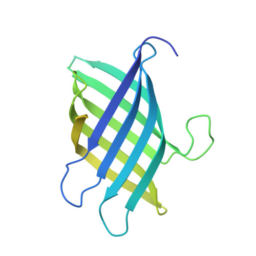
Macromolecular crystallography generally requires the recovery of missing phase information from diffraction data to reconstruct an electron-density map of the crystallized molecule. Most recent structures have been solved using molecular replacement as a phasing method, requiring an a priori structure that is closely related to the target protein to serve as a search model; when no such search model exists, molecular replacement is not possible. New advances in computational machine-learning methods, however, have resulted in major advances in protein structure predictions from sequence information. Methods that generate predicted structural models of sufficient accuracy provide a powerful approach to molecular replacement. Taking advantage of these advances, AlphaFold predictions were applied to enable structure determination of a bacterial protein of unknown function (UniProtKB Q63NT7, NCBI locus BPSS0212) based on diffraction data that had evaded phasing attempts using MIR and anomalous scattering methods. Using both X-ray and micro-electron (microED) diffraction data, it was possible to solve the structure of the main fragment of the protein using a predicted model of that domain as a starting point. The use of predicted structural models importantly expands the promise of electron diffraction, where structure determination relies critically on molecular replacement.
- Molecular Biology Institute, University of California, Los Angeles, Los Angeles, CA 90095, USA.
Organizational Affiliation: