8SZT

Structure of Kdac1 from Acinetobacter baumannii


Experimental Data Snapshot

  • Method: X-RAY DIFFRACTION
  • Resolution: 2.50 Å
  • R-Value Free: 0.262 
  • R-Value Work: 0.201 
  • R-Value Observed: 0.203 

wwPDB Validation   3D Report Full Report


This is version 1.1 of the entry. See complete history


Literature

Structure and Function of Kdac1, a Class II Deacetylase from the Multidrug-Resistant Pathogen Acinetobacter baumannii .

Watson, P.R.Christianson, D.W.

(2023) Biochemistry 62: 2689-2699

  • DOI: https://doi.org/10.1021/acs.biochem.3c00288
  • Primary Citation of Related Structures:  
    8SZT, 8SZU

  • PubMed Abstract: 

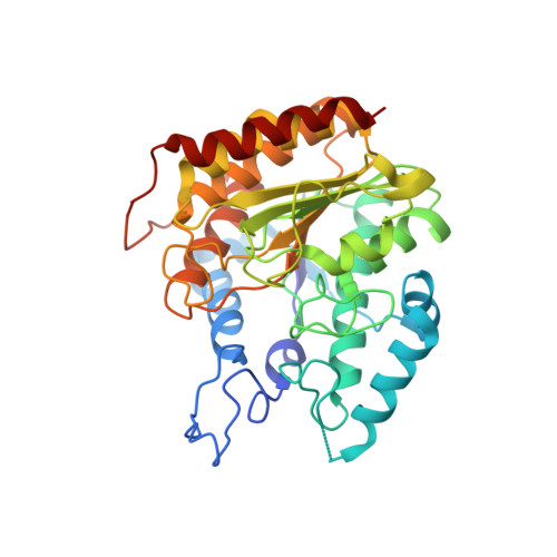
    Proteomics studies indicate that 10% of proteins in the opportunistic pathogen Acinetobacter baumannii are acetylated, suggesting that lysine acetyltransferases and deacetylases function to maintain and regulate a robust bacterial acetylome. As the first step in exploring these fascinating prokaryotic enzymes, we now report the preparation and characterization of the lysine deacetylase Kdac1. We show that Kdac1 catalyzes the deacetylation of free acetyllysine and acetyllysine tetrapeptide assay substrates, and we also report the X-ray crystal structures of unliganded Kdac1 as well as its complex with the hydroxamate inhibitor Citarinostat. Kdac1 is a tetramer in solution and in the crystal; the crystal structure reveals that the L1 loop functions to stabilize quaternary structure, forming inter-subunit hydrogen bonds and salt bridges around a central arginine residue (R30). Surprisingly, the L1 loop partially blocks entry to the active site, but it is sufficiently flexible to allow for the binding of two Citarinostat molecules in the active site. The L12 loop is also important for maintaining quaternary structure; here, a conserved arginine (R278) accepts hydrogen bonds from the backbone carbonyl groups of residues in an adjacent monomer. Structural comparisons with two other prokaryotic lysine deacetylases reveal conserved residues in the L1 and L12 loops that similarly support tetramer assembly. These studies provide a structural foundation for understanding enzymes that regulate protein function in bacteria through reversible lysine acetylation, serving as a first step in the exploration of these enzymes as possible targets for the development of new antibiotics.


  • Organizational Affiliation

    Roy and Diana Vagelos Laboratories, Department of Chemistry, University of Pennsylvania, 231 South 34th Street, Philadelphia, Pennsylvania 19104-6323, United States.


Macromolecules
Find similar proteins by:  (by identity cutoff)  |  3D Structure
Entity ID: 1
MoleculeChains Sequence LengthOrganismDetailsImage
Histone deacetylase domain-containing proteinA [auth B],
B [auth A],
C,
D
372Acinetobacter baumanniiMutation(s): 0 
UniProt
Find proteins for R8YPJ7 (Acinetobacter pittii ANC 4050)
Explore R8YPJ7 
Go to UniProtKB:  R8YPJ7
Entity Groups  
Sequence Clusters30% Identity50% Identity70% Identity90% Identity95% Identity100% Identity
UniProt GroupR8YPJ7
Sequence Annotations
Expand
  • Reference Sequence
Small Molecules
Ligands 3 Unique
IDChains Name / Formula / InChI Key2D Diagram3D Interactions
ZN
Query on ZN

Download Ideal Coordinates CCD File 
E [auth B],
I [auth A],
M [auth C],
Q [auth D]
ZINC ION
Zn
PTFCDOFLOPIGGS-UHFFFAOYSA-N
K
Query on K

Download Ideal Coordinates CCD File 
F [auth B]
G [auth B]
J [auth A]
K [auth A]
N [auth C]
F [auth B],
G [auth B],
J [auth A],
K [auth A],
N [auth C],
O [auth C],
R [auth D],
S [auth D]
POTASSIUM ION
K
NPYPAHLBTDXSSS-UHFFFAOYSA-N
CL
Query on CL

Download Ideal Coordinates CCD File 
H [auth B],
L [auth A],
P [auth C],
T [auth D]
CHLORIDE ION
Cl
VEXZGXHMUGYJMC-UHFFFAOYSA-M
Experimental Data & Validation

Experimental Data

  • Method: X-RAY DIFFRACTION
  • Resolution: 2.50 Å
  • R-Value Free: 0.262 
  • R-Value Work: 0.201 
  • R-Value Observed: 0.203 
  • Space Group: C 2 2 21
Unit Cell:
Length ( Å )Angle ( ˚ )
a = 113.949α = 90
b = 181.689β = 90
c = 155.33γ = 90
Software Package:
Software NamePurpose
PHENIXrefinement
iMOSFLMdata reduction
Aimlessdata scaling
PHASERphasing

Structure Validation

View Full Validation Report


Entry History & Funding Information

Deposition Data


Funding OrganizationLocationGrant Number
National Institutes of Health/National Institute of General Medical Sciences (NIH/NIGMS)United StatesGM49758

Revision History  (Full details and data files)

  • Version 1.0: 2023-09-06
    Type: Initial release
  • Version 1.1: 2023-10-04
    Changes: Database references