


Crystal Structure of Fluorescent Protein Fusion Red 2
Pletnev, S., Pletneva, N., Pletnev, V.To be published.
Experimental Data Snapshot
Starting Model: experimental
View more details
wwPDB Validation 3D Report Full Report

Entity ID: 1 | |||||
|---|---|---|---|---|---|
| Molecule | Chains | Sequence Length | Organism | Details | Image |
| Far-red Fluorescent Protein Fusion Red 2 | 243 | Entacmaea quadricolor | Mutation(s): 0 | 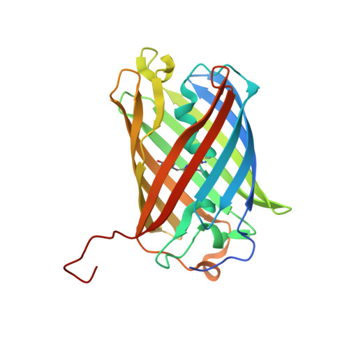 | |
UniProt | |||||
Entity Groups | |||||
| Sequence Clusters | 30% Identity50% Identity70% Identity90% Identity95% Identity100% Identity | ||||
| UniProt Group | D0VX33 | ||||
Sequence AnnotationsExpand | |||||
Reference Sequence | |||||
| Ligands 3 Unique | |||||
|---|---|---|---|---|---|
| ID | Chains | Name / Formula / InChI Key | 2D Diagram | 3D Interactions | |
| P6G Download:Ideal Coordinates CCD File | B [auth A] | HEXAETHYLENE GLYCOL C12 H26 O7 IIRDTKBZINWQAW-UHFFFAOYSA-N |  | ||
| GOL Download:Ideal Coordinates CCD File | C [auth A], D [auth A] | GLYCEROL C3 H8 O3 PEDCQBHIVMGVHV-UHFFFAOYSA-N |  | ||
| NO3 Download:Ideal Coordinates CCD File | E [auth A], F [auth A] | NITRATE ION N O3 NHNBFGGVMKEFGY-UHFFFAOYSA-N |  | ||
| Modified Residues 1 Unique | |||||
|---|---|---|---|---|---|
| ID | Chains | Type | Formula | 2D Diagram | Parent |
| NRQ Query on NRQ | A | L-PEPTIDE LINKING | C16 H17 N3 O4 S |  | MET, TYR, GLY |
| Length ( Å ) | Angle ( ˚ ) |
|---|---|
| a = 71.155 | α = 90 |
| b = 81.442 | β = 90 |
| c = 42.539 | γ = 90 |
| Software Name | Purpose |
|---|---|
| REFMAC | refinement |
| SCALEPACK | data scaling |
| HKL-2000 | data reduction |
| MOLREP | phasing |
| Funding Organization | Location | Grant Number |
|---|---|---|
| Russian Science Foundation | Russian Federation | 23-24-00011 |


