Structural determination and characterisation of the CYP105Q4 cytochrome P450 enzyme from Mycobacterium marinum.
Mohamed, H., Child, S.A., Doherty, D.Z., Bruning, J.B., Bell, S.G.(2024) Arch Biochem Biophys 754: 109950-109950
- PubMed: 38430969 Search on PubMed
- DOI: https://doi.org/10.1016/j.abb.2024.109950
- Primary Citation Related Structures:
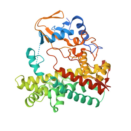
8SWL - PubMed Abstract:
The cytochrome P450 family of heme metalloenzymes (CYPs) catalyse important biological monooxygenation reactions. Mycobacterium marinum contains a gene encoding a CYP105Q4 enzyme of unknown function. Other members of the CYP105 CYP family have key roles in bacterial metabolism including the synthesis of secondary metabolites. We produced and purified the cytochrome P450 enzyme CYP105Q4 to enable its characterization. Several nitrogen-donor atom-containing ligands were found to bind to CYP105Q4 generating type II changes in the UV-vis absorbance spectrum. Based on the UV-vis absorbance spectra none of the potential substrate ligands we tested with CYP105Q4 were able to displace the sixth distal aqua ligand from the heme, though there was evidence for binding of oleic acid and amphotericin B. The crystal structure of CYP105Q4 in the substrate-free form was determined in an open conformation. A computational structural similarity search (Dali) was used to find the most closely related characterized relatives within the CYP105 family. The structure of CYP105Q4 enzyme was compared to the GfsF CYP enzyme from Streptomyces graminofaciens which is involved in the biosynthesis of a macrolide polyketide. This structural comparison to GfsF revealed conformational changes in the helices and loops near the entrance to the substrate access channel. A disordered B/C loop region, usually involved in substrate recognition, was also observed.
- Department of Chemistry, University of Adelaide, SA, 5005, Australia.
Organizational Affiliation: