8ST7

Structure of E3 ligase VsHECT bound to ubiquitin


Experimental Data Snapshot

  • Method: X-RAY DIFFRACTION
  • Resolution: 1.44 Å
  • R-Value Free: 0.199 
  • R-Value Work: 0.170 
  • R-Value Observed: 0.171 

wwPDB Validation   3D Report Full Report


This is version 1.2 of the entry. See complete history


Literature

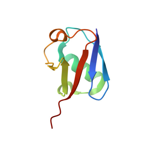
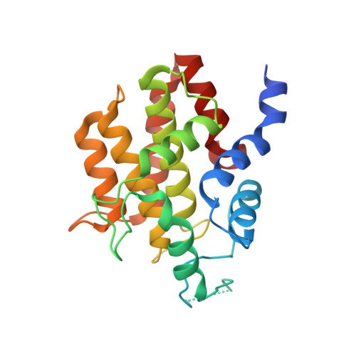
Bacterial ligases reveal fundamental principles of polyubiquitin specificity.

Franklin, T.G.Brzovic, P.S.Pruneda, J.N.

(2023) Mol Cell 83: 4538-4554.e4

  • DOI: https://doi.org/10.1016/j.molcel.2023.11.017
  • Primary Citation of Related Structures:  
    8ST7, 8ST8, 8ST9

  • PubMed Abstract: 

    Homologous to E6AP C terminus (HECT) E3 ubiquitin (Ub) ligases direct substrates toward distinct cellular fates dictated by the specific form of monomeric or polymeric Ub (polyUb) signal attached. How polyUb specificity is achieved has been a long-standing mystery, despite extensive study in various hosts, ranging from yeast to human. The bacterial pathogens enterohemorrhagic Escherichia coli and Salmonella Typhimurium encode outlying examples of "HECT-like" (bHECT) E3 ligases, but commonalities to eukaryotic HECT (eHECT) mechanism and specificity had not been explored. We expanded the bHECT family with examples in human and plant pathogens. Three bHECT structures in primed, Ub-loaded states resolved key details of the entire Ub ligation process. One structure provided a rare glimpse into the act of ligating polyUb, yielding a means to rewire polyUb specificity of both bHECT and eHECT ligases. Studying this evolutionarily distinct bHECT family has revealed insight into the function of key bacterial virulence factors as well as fundamental principles underlying HECT-type Ub ligation.


  • Organizational Affiliation

    Department of Molecular Microbiology and Immunology, Oregon Health and Science University, Portland, OR 97239, USA.


Macromolecules
Find similar proteins by:  (by identity cutoff)  |  3D Structure
Entity ID: 1
MoleculeChains Sequence LengthOrganismDetailsImage
UbiquitinA [auth D],
B
75Homo sapiensMutation(s): 0 
Gene Names: UBC
UniProt & NIH Common Fund Data Resources
Find proteins for P0CG48 (Homo sapiens)
Explore P0CG48 
Go to UniProtKB:  P0CG48
PHAROS:  P0CG48
GTEx:  ENSG00000150991 
Entity Groups  
Sequence Clusters30% Identity50% Identity70% Identity90% Identity95% Identity100% Identity
UniProt GroupP0CG48
Sequence Annotations
Expand
  • Reference Sequence
Find similar proteins by:  (by identity cutoff)  |  3D Structure
Entity ID: 2
MoleculeChains Sequence LengthOrganismDetailsImage
E3 ubiquitin-protein ligase SopA-like catalytic domain-containing proteinC [auth A],
D [auth C]
226VerrucomicrobiotaMutation(s): 0 
Gene Names: C5B47_04425
UniProt
Find proteins for A0A2V2RSR1 (Verrucomicrobiota bacterium)
Explore A0A2V2RSR1 
Go to UniProtKB:  A0A2V2RSR1
Entity Groups  
Sequence Clusters30% Identity50% Identity70% Identity90% Identity95% Identity100% Identity
UniProt GroupA0A2V2RSR1
Sequence Annotations
Expand
  • Reference Sequence
Experimental Data & Validation

Experimental Data

  • Method: X-RAY DIFFRACTION
  • Resolution: 1.44 Å
  • R-Value Free: 0.199 
  • R-Value Work: 0.170 
  • R-Value Observed: 0.171 
  • Space Group: P 1 21 1
Unit Cell:
Length ( Å )Angle ( ˚ )
a = 35.855α = 90
b = 157.276β = 93.756
c = 53.025γ = 90
Software Package:
Software NamePurpose
Web-Icedata collection
XDSdata reduction
Aimlessdata scaling
PHASERphasing
PHENIXrefinement

Structure Validation

View Full Validation Report


Entry History & Funding Information

Deposition Data


Funding OrganizationLocationGrant Number
National Institutes of Health/National Institute of General Medical Sciences (NIH/NIGMS)United StatesR35GM142486

Revision History  (Full details and data files)

  • Version 1.0: 2023-07-12
    Type: Initial release
  • Version 1.1: 2023-12-27
    Changes: Data collection, Database references
  • Version 1.2: 2024-01-03
    Changes: Database references