8SGE

KLHDC2 Kelch Domain with ligand KDRLKZ-1


Experimental Data Snapshot

  • Method: X-RAY DIFFRACTION
  • Resolution: 1.51 Å
  • R-Value Free: 0.206 
  • R-Value Work: 0.179 
  • R-Value Observed: 0.180 

wwPDB Validation   3D Report Full Report


Ligand Structure Quality Assessment 


This is version 1.2 of the entry. See complete history


Literature

Co-opting the E3 ligase KLHDC2 for targeted protein degradation by small molecules.

Hickey, C.M.Digianantonio, K.M.Zimmermann, K.Harbin, A.Quinn, C.Patel, A.Gareiss, P.Chapman, A.Tiberi, B.Dobrodziej, J.Corradi, J.Cacace, A.M.Langley, D.R.Bekes, M.

(2024) Nat Struct Mol Biol 31: 311-322

  • DOI: https://doi.org/10.1038/s41594-023-01146-w
  • Primary Citation of Related Structures:  
    8SGE, 8SGF, 8SH2

  • PubMed Abstract: 

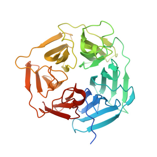
    Targeted protein degradation (TPD) by PROTAC (proteolysis-targeting chimera) and molecular glue small molecules is an emerging therapeutic strategy. To expand the roster of E3 ligases that can be utilized for TPD, we describe the discovery and biochemical characterization of small-molecule ligands targeting the E3 ligase KLHDC2. Furthermore, we functionalize these KLHDC2-targeting ligands into KLHDC2-based BET-family and AR PROTAC degraders and demonstrate KLHDC2-dependent target-protein degradation. Additionally, we offer insight into the assembly of the KLHDC2 E3 ligase complex. Using biochemical binding studies, X-ray crystallography and cryo-EM, we show that the KLHDC2 E3 ligase assembles into a dynamic tetramer held together via its own C terminus, and that this assembly can be modulated by substrate and ligand engagement.


  • Organizational Affiliation

    Arvinas, Inc, New Haven, CT, USA.


Macromolecules
Find similar proteins by:  (by identity cutoff)  |  3D Structure
Entity ID: 1
MoleculeChains Sequence LengthOrganismDetailsImage
Kelch domain-containing protein 2
A, B
362Homo sapiensMutation(s): 0 
Gene Names: KLHDC2HCA33
UniProt & NIH Common Fund Data Resources
Find proteins for Q9Y2U9 (Homo sapiens)
Explore Q9Y2U9 
Go to UniProtKB:  Q9Y2U9
PHAROS:  Q9Y2U9
GTEx:  ENSG00000165516 
Entity Groups  
Sequence Clusters30% Identity50% Identity70% Identity90% Identity95% Identity100% Identity
UniProt GroupQ9Y2U9
Sequence Annotations
Expand
  • Reference Sequence
Small Molecules
Ligands 2 Unique
IDChains Name / Formula / InChI Key2D Diagram3D Interactions
ZWS (Subject of Investigation/LOI)
Query on ZWS

Download Ideal Coordinates CCD File 
H [auth A],
M [auth B]
[(5P)-5-{3-[(2R)-butan-2-yl]-7-[(2-methoxyethoxy)carbonyl]-2-oxo-5,6,7,8-tetrahydro-1,7-naphthyridin-1(2H)-yl}-2-oxopyridin-1(2H)-yl]acetic acid
C23 H29 N3 O7
XFTKAVIYFGOXSI-OAHLLOKOSA-N
GOL
Query on GOL

Download Ideal Coordinates CCD File 
C [auth A]
D [auth A]
E [auth A]
F [auth A]
G [auth A]
C [auth A],
D [auth A],
E [auth A],
F [auth A],
G [auth A],
I [auth B],
J [auth B],
K [auth B],
L [auth B]
GLYCEROL
C3 H8 O3
PEDCQBHIVMGVHV-UHFFFAOYSA-N
Experimental Data & Validation

Experimental Data

  • Method: X-RAY DIFFRACTION
  • Resolution: 1.51 Å
  • R-Value Free: 0.206 
  • R-Value Work: 0.179 
  • R-Value Observed: 0.180 
  • Space Group: P 1 21 1
Unit Cell:
Length ( Å )Angle ( ˚ )
a = 44.9α = 90
b = 88.49β = 104.45
c = 89.22γ = 90
Software Package:
Software NamePurpose
BUSTERrefinement
autoPROCdata reduction
autoPROCdata scaling
PHASERphasing

Structure Validation

View Full Validation Report


Ligand Structure Quality Assessment 


Entry History & Funding Information

Deposition Data


Funding OrganizationLocationGrant Number
Other privateUnited States--

Revision History  (Full details and data files)

  • Version 1.0: 2024-01-03
    Type: Initial release
  • Version 1.1: 2024-01-17
    Changes: Database references
  • Version 1.2: 2024-02-28
    Changes: Database references