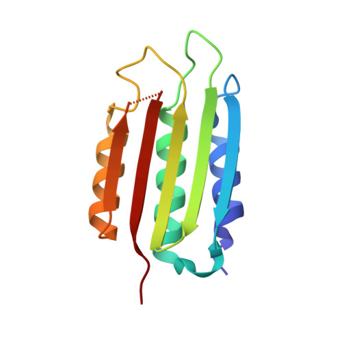
8SBM

Crystal structure of the wild-type Catalytic ATP-binding domain of Mtb DosS


Experimental Data Snapshot

  • Method: X-RAY DIFFRACTION
  • Resolution: 1.47 Å
  • R-Value Free: 0.193 
  • R-Value Work: 0.153 
  • R-Value Observed: 0.155 

wwPDB Validation   3D Report Full Report


This is version 1.1 of the entry. See complete history


Literature

Understanding ATP Binding to DosS Catalytic Domain with a Short ATP-Lid.

Larson, G.W.Windsor, P.K.Smithwick, E.Shi, K.Aihara, H.Rama Damodaran, A.Bhagi-Damodaran, A.

(2023) Biochemistry 62: 3283-3292

  • DOI: https://doi.org/10.1021/acs.biochem.3c00306
  • Primary Citation of Related Structures:  
    8SBM

  • PubMed Abstract: 

    DosS is a heme-containing histidine kinase that triggers dormancy transformation in Mycobacterium tuberculosis . Sequence comparison of the catalytic ATP-binding (CA) domain of DosS to other well-studied histidine kinases reveals a short ATP-lid. This feature has been thought to block binding of ATP to DosS's CA domain in the absence of interactions with DosS's dimerization and histidine phospho-transfer (DHp) domain. Here, we use a combination of computational modeling, structural biology, and biophysical studies to re-examine ATP-binding modalities in DosS. We show that the closed-lid conformation observed in crystal structures of DosS CA is caused by the presence of Zn 2+ in the ATP binding pocket that coordinates with Glu537 on the ATP-lid. Furthermore, circular dichroism studies and comparisons of DosS CA's crystal structure with its AlphaFold model and homologous DesK reveal that residues 503-507 that appear as a random coil in the Zn 2+ -coordinated crystal structure are in fact part of the N-box α helix needed for efficient ATP binding. Such random-coil transformation of an N-box α helix turn and the closed-lid conformation are both artifacts arising from large millimolar Zn 2+ concentrations used in DosS CA crystallization buffers. In contrast, in the absence of Zn 2+ , the short ATP-lid of DosS CA has significant conformational flexibility and can effectively bind AMP-PNP ( K d = 53 ± 13 μM), a non-hydrolyzable ATP analog. Furthermore, the nucleotide affinity remains unchanged when CA is conjugated to the DHp domain ( K d = 51 ± 6 μM). In all, our findings reveal that the short ATP-lid of DosS CA does not hinder ATP binding and provide insights that extend to 2988 homologous bacterial proteins containing such ATP-lids.


  • Organizational Affiliation

    Department of Chemistry, University of Minnesota, Minneapolis, Minnesota 55455, United States.


Macromolecules
Find similar proteins by:  (by identity cutoff)  |  3D Structure
Entity ID: 1
MoleculeChains Sequence LengthOrganismDetailsImage
GAF domain-containing protein
A, B
126Mycobacterium tuberculosisMutation(s): 0 
Gene Names: GJE03_16455
UniProt
Find proteins for P9WGK3 (Mycobacterium tuberculosis (strain ATCC 25618 / H37Rv))
Explore P9WGK3 
Go to UniProtKB:  P9WGK3
Entity Groups  
Sequence Clusters30% Identity50% Identity70% Identity90% Identity95% Identity100% Identity
UniProt GroupP9WGK3
Sequence Annotations
Expand
  • Reference Sequence
Small Molecules
Ligands 3 Unique
IDChains Name / Formula / InChI Key2D Diagram3D Interactions
ZN
Query on ZN

Download Ideal Coordinates CCD File 
C [auth A],
H [auth A],
J [auth B],
N [auth B]
ZINC ION
Zn
PTFCDOFLOPIGGS-UHFFFAOYSA-N
EDO
Query on EDO

Download Ideal Coordinates CCD File 
D [auth A]
E [auth A]
F [auth A]
G [auth A]
I [auth A]
D [auth A],
E [auth A],
F [auth A],
G [auth A],
I [auth A],
K [auth B],
L [auth B],
M [auth B]
1,2-ETHANEDIOL
C2 H6 O2
LYCAIKOWRPUZTN-UHFFFAOYSA-N
NA
Query on NA

Download Ideal Coordinates CCD File 
O [auth B]SODIUM ION
Na
FKNQFGJONOIPTF-UHFFFAOYSA-N
Experimental Data & Validation

Experimental Data

  • Method: X-RAY DIFFRACTION
  • Resolution: 1.47 Å
  • R-Value Free: 0.193 
  • R-Value Work: 0.153 
  • R-Value Observed: 0.155 
  • Space Group: C 1 2 1
Unit Cell:
Length ( Å )Angle ( ˚ )
a = 54.438α = 90
b = 61.775β = 107.54
c = 72.234γ = 90
Software Package:
Software NamePurpose
PHENIXrefinement
Aimlessdata scaling
PDB_EXTRACTdata extraction
XDSdata reduction
PHASERphasing

Structure Validation

View Full Validation Report


Entry History & Funding Information

Deposition Data


Funding OrganizationLocationGrant Number
National Institutes of Health/National Institute of General Medical Sciences (NIH/NIGMS)United StatesR35-GM118047

Revision History  (Full details and data files)

  • Version 1.0: 2023-11-08
    Type: Initial release
  • Version 1.1: 2023-11-29
    Changes: Database references