Comparative analysis of the physical properties of murine and human S100A7: Insight into why zinc piracy is mediated by human but not murine S100A7.
Harrison, S.A., Naretto, A., Balakrishnan, S., Perera, Y.R., Chazin, W.J.(2023) J Biological Chem 299: 105292-105292
- PubMed: 37769710 Search on PubMedSearch on PubMed Central
- DOI: https://doi.org/10.1016/j.jbc.2023.105292
- Primary Citation Related Structures:
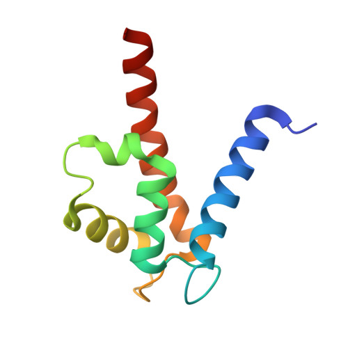
8S9W - PubMed Abstract:
S100 proteins are a subfamily of EF-hand calcium-binding proteins found primarily in vertebrate animals. They are distinguished by binding of transition metals and functioning in both the intracellular and extracellular milieu. S100A7 functions in the protection of the skin and mucous membranes and is a biomarker in inflammatory skin disease. A recent study of Neisseria gonorrhoeae infection revealed that human but not murine S100A7 could be used to evade host nutritional immunity. To understand the molecular basis for this difference, we carried out a comparative analysis of the physical and structural properties of human and murine S100A7. The X-ray crystal structure of Ca 2+ -loaded mouse S100A7 (mS100A7) was determined to 1.69 Å resolution, and Ca 2+ -induced conformational changes were assessed by NMR. Unlike human S100A7 (hS100A7), which exhibits conformational changes in response to binding of Ca 2+ , no significant changes in mS100A7 were detected. Dynamic light scattering, circular dichroism, and a competition chelator assay were used to compare the Zn 2+ affinity and the effects of ion binding on mS100A7 versus hS100A7. Alignment of their sequences revealed a substantial difference in the C-terminal region, which is an important mediator of protein-protein interactions, suggesting a rationale for the specificity of N. gonorrhoeae for hS100A7. These data, along with more detailed analysis of S100A7 sequence conservation across different species, support the proposal that, although hS100A7 is highly conserved in many mammals, the murine protein is a distinct ortholog. Our results highlight the potential limitations of using mouse models for studying bacterial infections in humans.
- Departments of Biochemistry and Chemistry, and Center for Structural Biology, Vanderbilt University, Nashville, Tennessee, USA.
Organizational Affiliation: