8S6A | pdb_00008s6a

X-ray structure of Dishevelled 3 PDZ domain in a complex with a class III peptide ligand


Experimental Data Snapshot

  • Method: X-RAY DIFFRACTION
  • Resolution: 1.36 Å
  • R-Value Free: 
    0.247 (Depositor), 0.245 (DCC) 
  • R-Value Work: 
    0.182 (Depositor), 0.189 (DCC) 
  • R-Value Observed: 
    0.185 (Depositor) 

Starting Model: experimental
View more details

wwPDB Validation   3D Report Full Report


This is version 1.2 of the entry. See complete history


Literature

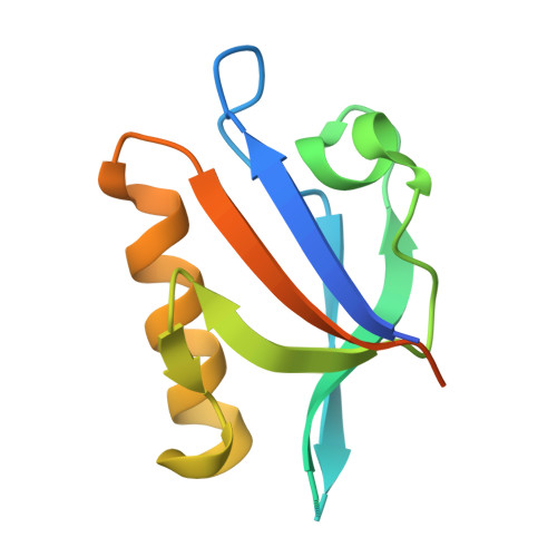
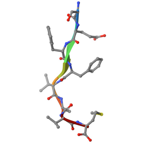
A class III ligand oscillates between internal and terminal binding modes as it engages with the Dishevelled PDZ domain.

Kumar, J.Micka, M.Komarek, J.Klumpler, T.Bystry, V.Sprangers, R.Barinka, C.Bryja, V.Tripsianes, K.

(2025) Structure 33: 1362

  • DOI: https://doi.org/10.1016/j.str.2025.05.012
  • Primary Citation of Related Structures:  
    8S6A

  • PubMed Abstract: 

    One of the largest domain-motif interactomes in human involves PSD-95/Discs-large/ZO-1 (PDZ) domains. The framework for understanding the PDZ interactome is well established; however the functional dynamics associated with PDZ-ligand interactions are poorly understood. Here, we report a dual PDZ-binding mode that ascribes unique dynamic features to class III ligand recognition. The crystal structure revealed that the PDZ domain can recognize either of the carboxylate moieties (terminal or internal) present in the class III ligand and laid out the register rules responsible for the dual recognition. Variants of the ligand designed to retain one or the other carboxylate of the native sequence were sufficient for PDZ binding. The conformational dynamics of PDZ probed by NMR relaxation dispersion experiments demonstrated that the class III ligand is shuffling binding modes as it engages with the PDZ domain. Our mechanistic findings reveal yet another aspect of PDZ binding plasticity specific to class III ligands.


  • Organizational Affiliation
    • CEITEC - Central European Institute of Technology, Masaryk University, Brno, Czech Republic.

Macromolecules
Find similar proteins by:  (by identity cutoff)  |  3D Structure
Entity ID: 1
MoleculeChains Sequence LengthOrganismDetailsImage
Segment polarity protein dishevelled homolog DVL-3A [auth B],
B [auth A]
111Homo sapiensMutation(s): 0 
Gene Names: DVL3KIAA0208
UniProt & NIH Common Fund Data Resources
Find proteins for Q92997 (Homo sapiens)
Explore Q92997 
Go to UniProtKB:  Q92997
PHAROS:  Q92997
GTEx:  ENSG00000161202 
Entity Groups  
Sequence Clusters30% Identity50% Identity70% Identity90% Identity95% Identity100% Identity
UniProt GroupQ92997
Sequence Annotations
Expand
  • Reference Sequence

Find similar proteins by:  Sequence   |   3D Structure  

Entity ID: 2
MoleculeChains Sequence LengthOrganismDetailsImage
C8 peptideC [auth P]8Homo sapiensMutation(s): 0 
Sequence Annotations
Expand
  • Reference Sequence
Experimental Data & Validation

Experimental Data

  • Method: X-RAY DIFFRACTION
  • Resolution: 1.36 Å
  • R-Value Free:  0.247 (Depositor), 0.245 (DCC) 
  • R-Value Work:  0.182 (Depositor), 0.189 (DCC) 
  • R-Value Observed: 0.185 (Depositor) 
Space Group: P 1 21 1
Unit Cell:
Length ( Å )Angle ( ˚ )
a = 46.91α = 90
b = 42.56β = 96.09
c = 48.29γ = 90
Software Package:
Software NamePurpose
REFMACrefinement
XDSdata reduction
Aimlessdata scaling
PHASERphasing

Structure Validation

View Full Validation Report


Entry History & Funding Information

Deposition Data


Funding OrganizationLocationGrant Number
Czech Academy of SciencesCzech Republic86652036

Revision History  (Full details and data files)

  • Version 1.0: 2025-03-12
    Type: Initial release
  • Version 1.1: 2025-06-25
    Changes: Database references
  • Version 1.2: 2025-08-20
    Changes: Database references