8RRY | pdb_00008rry

Crystal structure of copper-loaded SmAA10


Experimental Data Snapshot

  • Method: X-RAY DIFFRACTION
  • Resolution: 1.46 Å
  • R-Value Free: 
    0.188 (Depositor), 0.188 (DCC) 
  • R-Value Work: 
    0.160 (Depositor), 0.161 (DCC) 
  • R-Value Observed: 
    0.162 (Depositor) 

Starting Model: experimental
View more details

wwPDB Validation 3D Report Full Report

Validation slider image for 8RRY

Ligand Structure Quality Assessment 


This is version 1.2 of the entry. See complete history

Literature

Integrated Experimental and Theoretical Investigation of Copper Active Site Properties of a Lytic Polysaccharide Monooxygenase from Serratia marcescens.

Munzone, A.Pujol, M.Tamhankar, A.Joseph, C.Mazurenko, I.Reglier, M.Jannuzzi, S.A.V.Royant, A.Sicoli, G.DeBeer, S.Orio, M.Simaan, A.J.Decroos, C.

(2024) Inorg Chem 63: 11063-11078

  • DOI: https://doi.org/10.1021/acs.inorgchem.4c00602
  • Primary Citation Related Structures: 
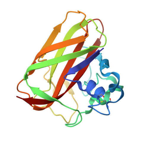
    8RRY

  • PubMed Abstract: 

    In this paper, we employed a multidisciplinary approach, combining experimental techniques and density functional theory (DFT) calculations to elucidate key features of the copper coordination environment of the bacterial lytic polysaccharide monooxygenase (LPMO) from Serratia marcescens ( Sm AA10). The structure of the holo -enzyme was successfully obtained by X-ray crystallography. We then determined the copper(II) binding affinity using competing ligands and observed that the affinity of the histidine brace ligands for copper is significantly higher than previously described. UV-vis, advanced electron paramagnetic resonance (EPR), and X-ray absorption spectroscopy (XAS) techniques, including high-energy resolution fluorescence detected (HERFD) XAS, were further used to gain insight into the copper environment in both the Cu(II) and Cu(I) redox states. The experimental data were successfully rationalized by DFT models, offering valuable information on the electronic structure and coordination geometry of the copper center. Finally, the Cu(II)/Cu(I) redox potential was determined using two different methods at ca . 350 mV vs NHE and rationalized by DFT calculations. This integrated approach not only advances our knowledge of the active site properties of Sm AA10 but also establishes a robust framework for future studies of similar enzymatic systems.


  • Organizational Affiliation
    • Aix Marseille Univ, CNRS, Centrale Méditerranée, iSm2, Marseille 13013, France.

Macromolecule Content 

  • Total Structure Weight: 38.32 kDa 
  • Atom Count: 3,224 
  • Modeled Residue Count: 340 
  • Deposited Residue Count: 340 
  • Unique protein chains: 1

Macromolecules

Find similar proteins by:|  3D Structure
Entity ID: 1
MoleculeChains  Sequence LengthOrganismDetailsImage
CBP21
A, B
170Serratia marcescensMutation(s): 0 
Gene Names: cbpgbpAAB868_00786BVG97_15155FOT62_12335HMI62_18715JBO39_14050NCTC10211_02621
EC: 1.14.99
UniProt
Find proteins for P9WEM3 (Serratia marcescens)
Explore P9WEM3 
Go to UniProtKB:  P9WEM3
Entity Groups
Sequence Clusters30% Identity50% Identity70% Identity90% Identity95% Identity100% Identity
UniProt GroupP9WEM3
Sequence Annotations
Expand
Reference Sequence

Small Molecules

Ligands 3 Unique
IDChains Name / Formula / InChI Key2D Diagram3D Interactions
CIT
(Subject of Investigation/LOI)

Query on CIT


Download:Ideal Coordinates CCD File
E [auth A],
H [auth B]
CITRIC ACID
C6 H8 O7
KRKNYBCHXYNGOX-UHFFFAOYSA-N
GOL
(Subject of Investigation/LOI)

Query on GOL


Download:Ideal Coordinates CCD File
D [auth A],
G [auth B]
GLYCEROL
C3 H8 O3
PEDCQBHIVMGVHV-UHFFFAOYSA-N
CU
(Subject of Investigation/LOI)

Query on CU


Download:Ideal Coordinates CCD File
C [auth A],
F [auth B]
COPPER (II) ION
Cu
JPVYNHNXODAKFH-UHFFFAOYSA-N

Experimental Data & Validation

Experimental Data

  • Method: X-RAY DIFFRACTION
  • Resolution: 1.46 Å
  • R-Value Free:  0.188 (Depositor), 0.188 (DCC) 
  • R-Value Work:  0.160 (Depositor), 0.161 (DCC) 
  • R-Value Observed: 0.162 (Depositor) 
Space Group: C 1 2 1
Unit Cell:
Length ( Å )Angle ( ˚ )
a = 53.646α = 90
b = 106.738β = 99.955
c = 55.459γ = 90
Software Package:
Software NamePurpose
PHENIXrefinement
PHENIXrefinement
XDSdata reduction
XSCALEdata scaling
PHASERphasing

Structure Validation

View Full Validation Report


Ligand Structure Quality Assessment 


Entry History 

& Funding Information

Deposition Data


Funding OrganizationLocationGrant Number
Agence Nationale de la Recherche (ANR)FranceANR-18-CE07-0003

Revision History  (Full details and data files)

  • Version 1.0: 2024-06-12
    Type: Initial release
  • Version 1.1: 2024-06-26
    Changes: Database references
  • Version 1.2: 2024-11-13
    Changes: Structure summary