8REX | pdb_00008rex

CryoEM structure of mouse GARP-lTGFbeta1 in complex with a Fab fragment derived from an activating antibody.


Experimental Data Snapshot

  • Method: ELECTRON MICROSCOPY
  • Resolution: 3.00 Å
  • Aggregation State: PARTICLE 
  • Reconstruction Method: SINGLE PARTICLE 

Starting Models: in silico
View more details

wwPDB Validation   3D Report Full Report


This is version 1.1 of the entry. See complete history


Literature

Antibody-mediated TGF-beta 1 activation for the treatment of diseases caused by deleterious T cell activity.

Lambert, F.Felix, J.Wautier, S.Dupre, E.Jamez, M.Michiels, C.Gaignage, M.Marien, L.Lesage, M.van der Woning, B.Savvides, S.N.Lucas, S.

(2025) Cell Rep 44: 116061-116061

  • DOI: https://doi.org/10.1016/j.celrep.2025.116061
  • Primary Citation of Related Structures:  
    8REW, 8REX

  • PubMed Abstract: 

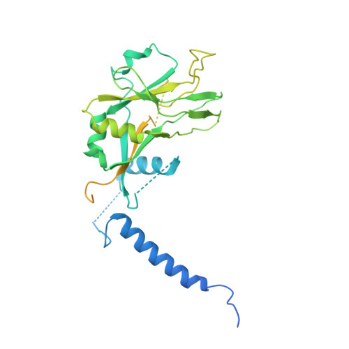
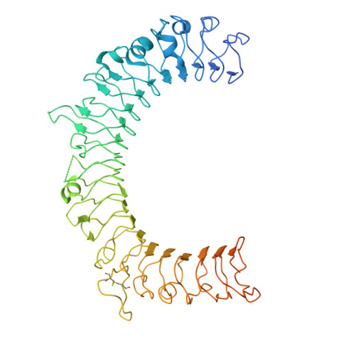
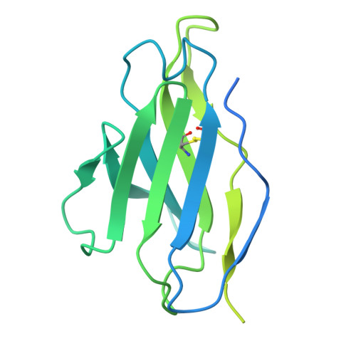
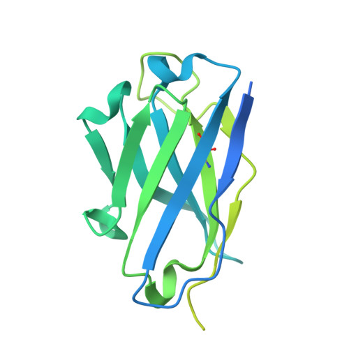
    Transforming growth factor β1 (TGF-β1) is an immunosuppressive cytokine produced as a latent homodimer, in which mature TGF-β1 is encapsulated and kept inactive by the latency-associated peptide (LAP). The transmembrane protein GARP presents latent TGF-β1 on the surface of regulatory T cells (Tregs) to enable activation and release of mature TGF-β1 by integrins. Here, we derived monoclonal antibodies (mAbs) that activate latent TGF-β1 anchored on cells by a transmembrane protein. Biochemical and structural studies by electron cryo-microscopy (cryo-EM) reveal that such mAb-mediated activation requires bivalent binding close to the LAP dimerization interface and crosslinking of two membrane-bound GARP:TGF-β1 complexes on the same cell or across different cells. Administration of mAbs to mice with graft versus host disease reduced disease severity and increased survival. The therapeutic effect required Tregs. Collectively, our findings demonstrate that activation of membrane-bound TGF-β1 in vivo is achievable with mAbs, introducing new immunotherapeutic options for allo- or autoimmune diseases characterized by deleterious T cell activity insufficiently controlled by Tregs.


  • Organizational Affiliation
    • de Duve Institute, UCLouvain, 1200 Brussels, Belgium.

Macromolecules
Find similar proteins by:  (by identity cutoff)  |  3D Structure
Entity ID: 1
MoleculeChains Sequence LengthOrganismDetailsImage
Transforming growth factor beta-1 proprotein
A, B, C, D
390Mus musculusMutation(s): 0 
Gene Names: Tgfb1
UniProt
Find proteins for P04202 (Mus musculus)
Explore P04202 
Go to UniProtKB:  P04202
Entity Groups  
Sequence Clusters30% Identity50% Identity70% Identity90% Identity95% Identity100% Identity
UniProt GroupP04202
Glycosylation
Glycosylation Sites: 1Go to GlyGen: P04202-1
Sequence Annotations
Expand
  • Reference Sequence
Find similar proteins by:  (by identity cutoff)  |  3D Structure
Entity ID: 2
MoleculeChains Sequence LengthOrganismDetailsImage
Transforming growth factor beta activator LRRC32675Mus musculusMutation(s): 0 
Gene Names: Lrrc32
UniProt
Find proteins for G3XA59 (Mus musculus)
Explore G3XA59 
Go to UniProtKB:  G3XA59
Entity Groups  
Sequence Clusters30% Identity50% Identity70% Identity90% Identity95% Identity100% Identity
UniProt GroupG3XA59
Glycosylation
Glycosylation Sites: 2Go to GlyGen: G3XA59-1
Sequence Annotations
Expand
  • Reference Sequence
Find similar proteins by:  (by identity cutoff)  |  3D Structure
Entity ID: 3
MoleculeChains Sequence LengthOrganismDetailsImage
mFab LMT-12, Light Chain
F, I
236Mus musculusMutation(s): 0 
Entity Groups  
Sequence Clusters30% Identity50% Identity70% Identity90% Identity95% Identity100% Identity
Sequence Annotations
Expand
  • Reference Sequence
Find similar proteins by:  (by identity cutoff)  |  3D Structure
Entity ID: 4
MoleculeChains Sequence LengthOrganismDetailsImage
mFab LMT-12, Heavy Chain
G, H
247Mus musculusMutation(s): 0 
Entity Groups  
Sequence Clusters30% Identity50% Identity70% Identity90% Identity95% Identity100% Identity
Sequence Annotations
Expand
  • Reference Sequence
Oligosaccharides

Help

Entity ID: 5
MoleculeChains Length2D Diagram Glycosylation3D Interactions
2-acetamido-2-deoxy-beta-D-glucopyranose-(1-4)-2-acetamido-2-deoxy-beta-D-glucopyranose
J, K, L
2N-Glycosylation
Glycosylation Resources
GlyTouCan:  G42666HT
GlyCosmos:  G42666HT
GlyGen:  G42666HT
Experimental Data & Validation

Experimental Data

  • Method: ELECTRON MICROSCOPY
  • Resolution: 3.00 Å
  • Aggregation State: PARTICLE 
  • Reconstruction Method: SINGLE PARTICLE 
EM Software:
TaskSoftware PackageVersion
RECONSTRUCTIONcryoSPARC
MODEL REFINEMENTPHENIX1.19.2-4158

Structure Validation

View Full Validation Report


Entry History & Funding Information

Deposition Data


Funding OrganizationLocationGrant Number
Other privateFondation contre le Cancer / 2020-079
Other governmentARC / 19/24-098
Fonds National de la Recherche Scientifique (FNRS)BelgiumPDR / T.0145.21
Fonds National de la Recherche Scientifique (FNRS)BelgiumWELBIO / CR2019A-02R
Other privateSalus Sanguinis

Revision History  (Full details and data files)

  • Version 1.0: 2025-06-25
    Type: Initial release
  • Version 1.1: 2025-10-08
    Changes: Data collection, Database references