Oligomerization mediated by the D2 domain of DTX3L is critical for DTX3L-PARP9 reading function of mono-ADP-ribosylated androgen receptor.
Vela-Rodriguez, C., Yang, C., Alanen, H.I., Eki, R., Abbas, T.A., Maksimainen, M.M., Glumoff, T., Duman, R., Wagner, A., Paschal, B.M., Lehtio, L.(2024) Protein Sci 33: e4945-e4945
- PubMed: 38511494 Search on PubMedSearch on PubMed Central
- DOI: https://doi.org/10.1002/pro.4945
- Primary Citation Related Structures:
8R79 - PubMed Abstract:
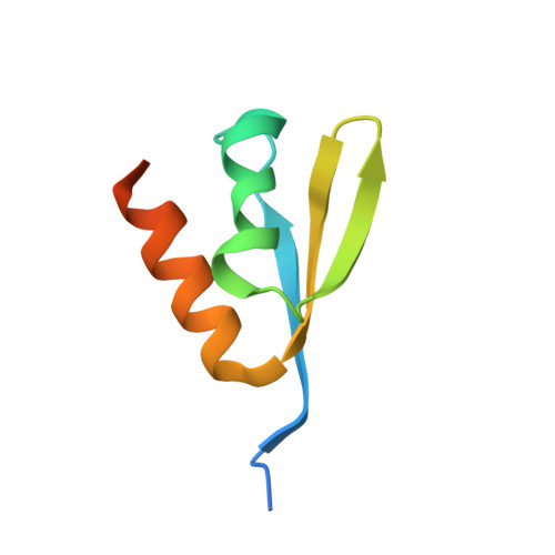
Deltex proteins are a family of E3 ubiquitin ligases that encode C-terminal RING and DTC domains that mediate interactions with E2 ubiquitin-conjugating enzymes and recognize ubiquitination substrates. DTX3L is unique among the Deltex proteins based on its N-terminal domain architecture. The N-terminal D1 and D2 domains of DTX3L mediate homo-oligomerization, and the D3 domain interacts with PARP9, a protein that contains tandem macrodomains with ADP-ribose reader function. While DTX3L and PARP9 are known to heterodimerize, and assemble into a high molecular weight oligomeric complex, the nature of the oligomeric structure, including whether this contributes to the ADP-ribose reader function is unknown. Here, we report a crystal structure of the DTX3L N-terminal D2 domain and show that it forms a tetramer with, conveniently, D2 symmetry. We identified two interfaces in the structure: a major, conserved interface with a surface of 973 Å 2 and a smaller one of 415 Å 2 . Using native mass spectrometry, we observed molecular species that correspond to monomers, dimers and tetramers of the D2 domain. Reconstitution of DTX3L knockout cells with a D1-D2 deletion mutant showed the domain is dispensable for DTX3L-PARP9 heterodimer formation, but necessary to assemble an oligomeric complex with efficient reader function for ADP-ribosylated androgen receptor. Our results suggest that homo-oligomerization of DTX3L is important for the DTX3L-PARP9 complex to read mono-ADP-ribosylation on a ligand-regulated transcription factor.
- Faculty of Biochemistry and Molecular Medicine & Biocenter Oulu, University of Oulu, Oulu, Finland.
Organizational Affiliation: