8QSR | pdb_00008qsr

Cryo-EM structure of the glucose-specific PTS transporter IICB from E. coli in the inward-facing conformation


Experimental Data Snapshot

  • Method: ELECTRON MICROSCOPY
  • Resolution: 2.56 Å
  • Aggregation State: PARTICLE 
  • Reconstruction Method: SINGLE PARTICLE 

Starting Model: in silico
View more details

wwPDB Validation   3D Report Full Report


This is version 1.1 of the entry. See complete history


Literature

Structure and mechanism of a phosphotransferase system glucose transporter.

Roth, P.Jeckelmann, J.M.Fender, I.Ucurum, Z.Lemmin, T.Fotiadis, D.

(2024) Nat Commun 15: 7992-7992

  • DOI: https://doi.org/10.1038/s41467-024-52100-3
  • Primary Citation of Related Structures:  
    8QSR, 8QST

  • PubMed Abstract: 

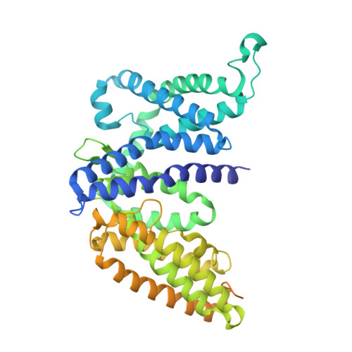
    Glucose is the primary source of energy for many organisms and is efficiently taken up by bacteria through a dedicated transport system that exhibits high specificity. In Escherichia coli, the glucose-specific transporter IICB Glc serves as the major glucose transporter and functions as a component of the phosphoenolpyruvate-dependent phosphotransferase system. Here, we report cryo-electron microscopy (cryo-EM) structures of the glucose-bound IICB Glc protein. The dimeric transporter embedded in lipid nanodiscs was captured in the occluded, inward- and occluded, outward-facing conformations. Together with biochemical and biophysical analyses, and molecular dynamics (MD) simulations, we provide insights into the molecular basis and dynamics for substrate recognition and binding, including the gates regulating the binding sites and their accessibility. By combination of these findings, we present a mechanism for glucose transport across the plasma membrane. Overall, this work provides molecular insights into the structure, dynamics, and mechanism of the IICB Glc transporter in a native-like lipid environment.


  • Organizational Affiliation

    Institute of Biochemistry and Molecular Medicine, Medical Faculty, University of Bern, Bern, Switzerland.


Macromolecules
Find similar proteins by:  (by identity cutoff)  |  3D Structure
Entity ID: 1
MoleculeChains Sequence LengthOrganismDetailsImage
PTS system glucose-specific EIICB component
A, B
499Escherichia coliMutation(s): 0 
Gene Names: ptsG
EC: 2.7.1.199
Membrane Entity: Yes 
UniProt
Find proteins for P69786 (Escherichia coli (strain K12))
Explore P69786 
Go to UniProtKB:  P69786
Entity Groups  
Sequence Clusters30% Identity50% Identity70% Identity90% Identity95% Identity100% Identity
UniProt GroupP69786
Sequence Annotations
Expand
  • Reference Sequence
Experimental Data & Validation

Experimental Data

  • Method: ELECTRON MICROSCOPY
  • Resolution: 2.56 Å
  • Aggregation State: PARTICLE 
  • Reconstruction Method: SINGLE PARTICLE 
EM Software:
TaskSoftware PackageVersion
RECONSTRUCTIONcryoSPARC3.2.0
MODEL REFINEMENTPHENIX

Structure Validation

View Full Validation Report


Entry History & Funding Information

Deposition Data


Funding OrganizationLocationGrant Number
Swiss National Science FoundationSwitzerland184980

Revision History  (Full details and data files)

  • Version 1.0: 2024-09-25
    Type: Initial release
  • Version 1.1: 2025-01-22
    Changes: Data collection, Database references, Structure summary