8QNS | pdb_00008qns

Crystal structure of murine AIF bound to N-terminal domain of CHCHD4


Experimental Data Snapshot

  • Method: X-RAY DIFFRACTION
  • Resolution: 3.21 Å
  • R-Value Free: 
    0.284 (Depositor), 0.279 (DCC) 
  • R-Value Work: 
    0.233 (Depositor), 0.230 (DCC) 

Starting Model: experimental
View more details

wwPDB Validation 3D Report Full Report

Validation slider image for 8QNS

Ligand Structure Quality Assessment 


This is version 1.1 of the entry. See complete history

Literature

CHCHD4 binding affects the active site of apoptosis inducing factor (AIF): Structural determinants for allosteric regulation.

Fagnani, E.Cocomazzi, P.Pellegrino, S.Tedeschi, G.Scalvini, F.G.Cossu, F.Da Vela, S.Aliverti, A.Mastrangelo, E.Milani, M.

(2024) Structure 32: 594-602.e4

  • DOI: https://doi.org/10.1016/j.str.2024.02.008
  • Primary Citation Related Structures: 
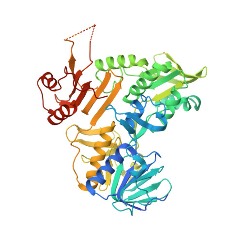
    8QNS

  • PubMed Abstract: 

    Apoptosis-inducing factor (AIF), which is confined to mitochondria of normal healthy cells, is the first identified caspase-independent cell death effector. Moreover, AIF is required for the optimal functioning of the respiratory chain machinery. Recent findings have revealed that AIF fulfills its pro-survival function by interacting with CHCHD4, a soluble mitochondrial protein which promotes the entrance and the oxidative folding of different proteins in the inner membrane space. Here, we report the crystal structure of the ternary complex involving the N-terminal 27-mer peptide of CHCHD4, NAD + , and AIF harboring its FAD (flavin adenine dinucleotide) prosthetic group in oxidized form. Combining this information with biophysical and biochemical data on the CHCHD4/AIF complex, we provide a detailed structural description of the interaction between the two proteins, validated by both chemical cross-linking mass spectrometry analysis and site-directed mutagenesis.


  • Organizational Affiliation
    • Biophysics Institute, CNR-IBF, Via Corti 12, 20133 Milan, Italy; Department of Bioscience, Università degli Studi di Milano, Via Celoria 26, 20133 Milan, Italy.

Macromolecule Content 

  • Total Structure Weight: 247.05 kDa 
  • Atom Count: 14,701 
  • Modeled Residue Count: 1,832 
  • Deposited Residue Count: 2,204 
  • Unique protein chains: 2

Macromolecules

Find similar proteins by:|  3D Structure
Entity ID: 1
MoleculeChains  Sequence LengthOrganismDetailsImage
Apoptosis-inducing factor 1, mitochondrialA,
B [auth D],
C [auth G],
D [auth J]
524Mus musculusMutation(s): 0 
Gene Names: Aifm1AifPdcd8
EC: 1.6.99
UniProt
Find proteins for Q9Z0X1 (Mus musculus)
Explore Q9Z0X1 
Go to UniProtKB:  Q9Z0X1
Entity Groups
Sequence Clusters30% Identity50% Identity70% Identity90% Identity95% Identity100% Identity
UniProt GroupQ9Z0X1
Sequence Annotations
Expand
Reference Sequence
Find similar proteins by:|  3D Structure
Entity ID: 2
MoleculeChains  Sequence LengthOrganismDetailsImage
Mitochondrial intermembrane space import and assembly protein 40E [auth M],
F [auth N],
G [auth O],
H [auth P]
27Mus musculusMutation(s): 0 
UniProt
Find proteins for Q8VEA4 (Mus musculus)
Explore Q8VEA4 
Go to UniProtKB:  Q8VEA4
Entity Groups
Sequence Clusters30% Identity50% Identity70% Identity90% Identity95% Identity100% Identity
UniProt GroupQ8VEA4
Sequence Annotations
Expand
Reference Sequence

Small Molecules

Ligands 2 Unique
IDChains Name / Formula / InChI Key2D Diagram3D Interactions
FAD

Query on FAD


Download:Ideal Coordinates CCD File
I [auth A],
K [auth D],
M [auth G],
O [auth J]
FLAVIN-ADENINE DINUCLEOTIDE
C27 H33 N9 O15 P2
VWWQXMAJTJZDQX-UYBVJOGSSA-N
NAD
(Subject of Investigation/LOI)

Query on NAD


Download:Ideal Coordinates CCD File
J [auth A],
L [auth D],
N [auth G],
P [auth J]
NICOTINAMIDE-ADENINE-DINUCLEOTIDE
C21 H27 N7 O14 P2
BAWFJGJZGIEFAR-NNYOXOHSSA-N

Experimental Data & Validation

Experimental Data

  • Method: X-RAY DIFFRACTION
  • Resolution: 3.21 Å
  • R-Value Free:  0.284 (Depositor), 0.279 (DCC) 
  • R-Value Work:  0.233 (Depositor), 0.230 (DCC) 
Space Group: P 21 21 21
Unit Cell:
Length ( Å )Angle ( ˚ )
a = 110.087α = 90
b = 115.619β = 90
c = 192.727γ = 90
Software Package:
Software NamePurpose
REFMACrefinement
XDSdata reduction
STARANISOdata scaling
MOLREPphasing

Structure Validation

View Full Validation Report


Ligand Structure Quality Assessment 


Entry History 

& Funding Information

Deposition Data


Funding OrganizationLocationGrant Number
European Union (EU)European Union--

Revision History  (Full details and data files)

  • Version 1.0: 2024-05-08
    Type: Initial release
  • Version 1.1: 2024-11-20
    Changes: Database references, Structure summary