8QH4

Crystal structure of reduced respiratory Complex I subunits NuoEF from Aquifex aeolicus bound to oxidized 3-acetylpyridine adenine dinucleotide


Experimental Data Snapshot

  • Method: X-RAY DIFFRACTION
  • Resolution: 1.96 Å
  • R-Value Free: 0.224 
  • R-Value Work: 0.192 

wwPDB Validation   3D Report Full Report


Ligand Structure Quality Assessment 


This is version 1.0 of the entry. See complete history


Literature

Structures of 3-acetylpyridine adenine dinucleotide and ADP-ribose bound to the electron input module of respiratory complex I.

Wohlwend, D.Merono, L.Bucka, S.Ritter, K.Jessen, H.J.Friedrich, T.

(2024) Structure 

  • DOI: https://doi.org/10.1016/j.str.2024.02.013
  • Primary Citation of Related Structures:  
    8QG1, 8QGW, 8QH4, 8QH7, 8QHK

  • PubMed Abstract: 

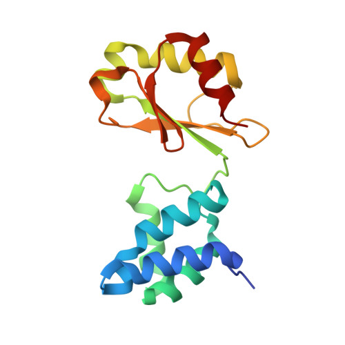
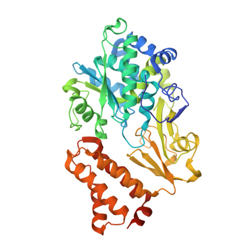
    Energy-converting NADH:ubiquinone oxidoreductase, respiratory complex I, is a major enzyme of energy metabolism that couples NADH oxidation and ubiquinone reduction with proton translocation. The NADH oxidation site features different enzymatic activities with various nucleotides. While the kinetics of these reactions are well described, only binding of NAD + and NADH have been structurally characterized. Here, we report the structures of the electron input module of Aquifex aeolicus complex I with bound ADP-ribose and 3-acetylpyridine adenine dinucleotides at resolutions better than 2.0 Å. ADP-ribose acts as inhibitor by blocking the "ADP-handle" motif essential for nucleotide binding. The pyridine group of APADH is minimally offset from flavin, which could contribute to its poorer suitability as substrate. A comparison with other nucleotide co-structures surprisingly shows that the adenine ribose and the pyrophosphate moiety contribute most to nucleotide binding, thus all adenine dinucleotides share core binding modes to the unique Rossmann-fold in complex I.


  • Organizational Affiliation

    Institute of Biochemistry, Albert-Ludwigs-Universität Freiburg, Freiburg, Germany.


Macromolecules
Find similar proteins by:  (by identity cutoff)  |  3D Structure
Entity ID: 1
MoleculeChains Sequence LengthOrganismDetailsImage
NADH-quinone oxidoreductase subunit E
A, C
160Aquifex aeolicus VF5Mutation(s): 0 
Gene Names: nuoEaq_574
EC: 7.1.1
UniProt
Find proteins for O66842 (Aquifex aeolicus (strain VF5))
Explore O66842 
Go to UniProtKB:  O66842
Entity Groups  
Sequence Clusters30% Identity50% Identity70% Identity90% Identity95% Identity100% Identity
UniProt GroupO66842
Sequence Annotations
Expand
  • Reference Sequence
Find similar proteins by:  (by identity cutoff)  |  3D Structure
Entity ID: 2
MoleculeChains Sequence LengthOrganismDetailsImage
NADH-quinone oxidoreductase subunit F
B, D
434Aquifex aeolicus VF5Mutation(s): 0 
Gene Names: nuoF
UniProt
Find proteins for O66841 (Aquifex aeolicus (strain VF5))
Explore O66841 
Go to UniProtKB:  O66841
Entity Groups  
Sequence Clusters30% Identity50% Identity70% Identity90% Identity95% Identity100% Identity
UniProt GroupO66841
Sequence Annotations
Expand
  • Reference Sequence
Small Molecules
Ligands 8 Unique
IDChains Name / Formula / InChI Key2D Diagram3D Interactions
A3D (Subject of Investigation/LOI)
Query on A3D

Download Ideal Coordinates CCD File 
J [auth B],
X [auth D]
3-ACETYLPYRIDINE ADENINE DINUCLEOTIDE
C22 H28 N6 O14 P2
KPVQNXLUPNWQHM-RBEMOOQDSA-N
FNR (Subject of Investigation/LOI)
Query on FNR

Download Ideal Coordinates CCD File 
I [auth B],
W [auth D]
1-DEOXY-1-(7,8-DIMETHYL-2,4-DIOXO-3,4-DIHYDRO-2H-BENZO[G]PTERIDIN-1-ID-10(5H)-YL)-5-O-PHOSPHONATO-D-RIBITOL
C17 H23 N4 O9 P
YTNIXZGTHTVJBW-SCRDCRAPSA-N
SF4 (Subject of Investigation/LOI)
Query on SF4

Download Ideal Coordinates CCD File 
H [auth B],
V [auth D]
IRON/SULFUR CLUSTER
Fe4 S4
LJBDFODJNLIPKO-UHFFFAOYSA-N
FES (Subject of Investigation/LOI)
Query on FES

Download Ideal Coordinates CCD File 
E [auth A],
R [auth C]
FE2/S2 (INORGANIC) CLUSTER
Fe2 S2
NIXDOXVAJZFRNF-UHFFFAOYSA-N
SO4
Query on SO4

Download Ideal Coordinates CCD File 
AA [auth D]
F [auth A]
G [auth A]
L [auth B]
S [auth C]
AA [auth D],
F [auth A],
G [auth A],
L [auth B],
S [auth C],
T [auth C],
Z [auth D]
SULFATE ION
O4 S
QAOWNCQODCNURD-UHFFFAOYSA-L
GOL
Query on GOL

Download Ideal Coordinates CCD File 
K [auth B],
Y [auth D]
GLYCEROL
C3 H8 O3
PEDCQBHIVMGVHV-UHFFFAOYSA-N
K
Query on K

Download Ideal Coordinates CCD File 
DA [auth D]POTASSIUM ION
K
NPYPAHLBTDXSSS-UHFFFAOYSA-N
NA
Query on NA

Download Ideal Coordinates CCD File 
BA [auth D]
CA [auth D]
M [auth B]
N [auth B]
O [auth B]
BA [auth D],
CA [auth D],
M [auth B],
N [auth B],
O [auth B],
P [auth B],
Q [auth B],
U [auth C]
SODIUM ION
Na
FKNQFGJONOIPTF-UHFFFAOYSA-N
Experimental Data & Validation

Experimental Data

  • Method: X-RAY DIFFRACTION
  • Resolution: 1.96 Å
  • R-Value Free: 0.224 
  • R-Value Work: 0.192 
  • Space Group: P 21 21 21
Unit Cell:
Length ( Å )Angle ( ˚ )
a = 63.527α = 90
b = 116.619β = 90
c = 190.01γ = 90
Software Package:
Software NamePurpose
REFMACrefinement
autoPROCdata reduction
Aimlessdata scaling
REFMACphasing

Structure Validation

View Full Validation Report


Ligand Structure Quality Assessment 


Entry History & Funding Information

Deposition Data


Funding OrganizationLocationGrant Number
Not funded--

Revision History  (Full details and data files)

  • Version 1.0: 2024-04-03
    Type: Initial release