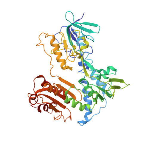
The crystal structure of mycothiol disulfide reductase (Mtr) provides mechanistic insight into the specific low-molecular-weight thiol reductase activity of Actinobacteria.
Gutierrez-Fernandez, J., Hersleth, H.P., Hammerstad, M.(2024) Acta Crystallogr D Struct Biol 80: 181-193
- PubMed: 38372589 Search on PubMedSearch on PubMed Central
- DOI: https://doi.org/10.1107/S205979832400113X
- Primary Citation Related Structures:
8QCJ, 8QCK - PubMed Abstract:
Low-molecular-weight (LMW) thiols are involved in many processes in all organisms, playing a protective role against reactive species, heavy metals, toxins and antibiotics. Actinobacteria, such as Mycobacterium tuberculosis, use the LMW thiol mycothiol (MSH) to buffer the intracellular redox environment. The NADPH-dependent FAD-containing oxidoreductase mycothiol disulfide reductase (Mtr) is known to reduce oxidized mycothiol disulfide (MSSM) to MSH, which is crucial to maintain the cellular redox balance. In this work, the first crystal structures of Mtr are presented, expanding the structural knowledge and understanding of LMW thiol reductases. The structural analyses and docking calculations provide insight into the nature of Mtrs, with regard to the binding and reduction of the MSSM substrate, in the context of related oxidoreductases. The putative binding site for MSSM suggests a similar binding to that described for the homologous glutathione reductase and its respective substrate glutathione disulfide, but with distinct structural differences shaped to fit the bulkier MSSM substrate, assigning Mtrs as uniquely functioning reductases. As MSH has been acknowledged as an attractive antitubercular target, the structural findings presented in this work may contribute towards future antituberculosis drug development.
- Section for Biochemistry and Molecular Biology, Department of Biosciences, University of Oslo, PO Box 1066, Blindern, 0316 Oslo, Norway.
Organizational Affiliation: