Structural Study of a New MbtI-Inhibitor Complex: Towards an Optimized Model for Structure-Based Drug Discovery.
Mori, M., Villa, S., Chiarelli, L.R., Meneghetti, F., Bellinzoni, M.(2023) Pharmaceuticals (Basel) 16
- PubMed: 38004425 Search on PubMedSearch on PubMed Central
- DOI: https://doi.org/10.3390/ph16111559
- Primary Citation Related Structures:
8QC4, 8QN5 - PubMed Abstract:
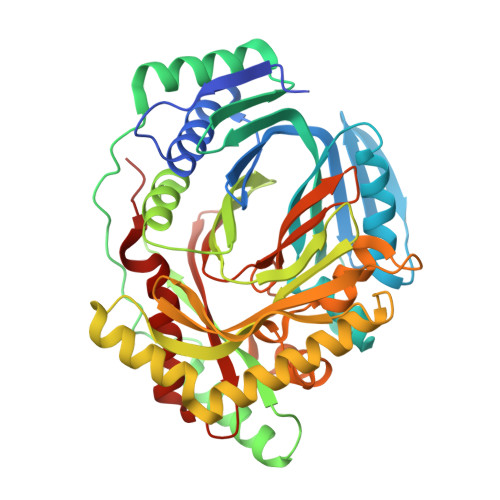
MbtI from Mycobacterium tuberculosis ( Mtb ) is a Mg 2+ -dependent salicylate synthase, belonging to the chorismate-utilizing enzyme (CUE) family. As a fundamental player in iron acquisition, MbtI promotes the survival and pathogenicity of Mtb in the infected host. Hence, it has emerged in the last decade as an innovative, potential target for the anti-virulence therapy of tuberculosis. In this context, 5-phenylfuran-2-carboxylic acids have been identified as potent MbtI inhibitors. The first co-crystal structure of MbtI in complex with a member of this class was described in 2020, showing the enzyme adopting an open configuration. Due to the high mobility of the loop adjacent to the binding pocket, large portions of the amino acid chain were not defined in the electron density map, hindering computational efforts aimed at structure-driven ligand optimization. Herein, we report a new, high-resolution co-crystal structure of MbtI with a furan-based derivative, in which the closed configuration of the enzyme allowed tracing the entirety of the active site pocket in the presence of the bound inhibitor. Moreover, we describe a new crystal structure of MbtI in open conformation and in complex with the known inhibitor methyl-AMT, suggesting that in vitro potency is not related to the observed enzyme conformation. These findings will prove fundamental to enhance the potency of this series via rational structure-based drug-design approaches.
- Department of Pharmaceutical Sciences, University of Milan, Via L. Mangiagalli 25, 20133 Milano, Italy.
Organizational Affiliation: