CI:Mor interactions in the lysogeny switches of Lactococcus lactis TP901-1 and Staphylococcus aureus phi 13 bacteriophages.
Varming, A.K., Huang, Z., Hamad, G.M., Rasmussen, K.K., Ingmer, H., Kilstrup, M., Lo Leggio, L.(2024) Microbiome Res Rep 3: 15-15
- PubMed: 38841409 Search on PubMedSearch on PubMed Central
- DOI: https://doi.org/10.20517/mrr.2023.50
- Primary Citation Related Structures:
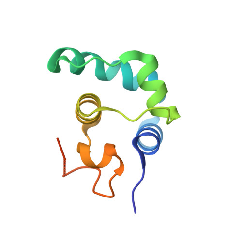
8QAO - PubMed Abstract:
Aim: To structurally characterize in detail the interactions between the phage repressor (CI) and the antirepressor (Mor) in the lysis-lysogeny switches of two Gram-positive bacteriophages, the lactococcal TP901-1 and staphylococcal φ13. Methods: We use crystallographic structure determination, computational structural modeling, and analysis, as well as biochemical methods, to elucidate similarities and differences in the CI:Mor interactions for the two genetic switches. Results: By comparing a newly determined and other available crystal structures for the N-terminal domain of CI (CI-NTD), we show that the CI interface involved in Mor binding undergoes structural changes upon binding in TP901-1. Most importantly, we show experimentally for the first time the direct interaction between CI and Mor for φ13, and model computationally the interaction interface. The computational modeling supports similar side chain rearrangements in TP901-1 and φ13. Conclusion: This study ascertains experimentally that, like in the TP901-1 lysogeny switch, staphylococcal φ13 CI and Mor interact with each other. The structural basis of the interaction of φ13 CI and Mor was computationally modeled and is similar to the interaction demonstrated experimentally between TP901-1 CI-NTD and Mor, likely involving similar rearrangement of residue side chains during the formation of the complex. The study identifies one CI residue, Glu69, which unusually interacts primarily through its aliphatic chain with an aromatic residue on Mor after changing its conformation compared to the un-complexed structure. This and other residues at the interface are suggested for investigation in future studies.
- Department of Chemistry, University of Copenhagen, Copenhagen DK-2100, Denmark.
Organizational Affiliation: