8Q2E | pdb_00008q2e

The 1.68-A X-ray crystal structure of Sporosarcina pasteurii urease inhibited by thiram and bound to dimethylditiocarbamate


Experimental Data Snapshot

  • Method: X-RAY DIFFRACTION
  • Resolution: 1.68 Å
  • R-Value Free: 
    0.163 (Depositor), 0.174 (DCC) 
  • R-Value Work: 
    0.138 (Depositor), 0.153 (DCC) 
  • R-Value Observed: 
    0.139 (Depositor) 

Starting Model: experimental
View more details

wwPDB Validation 3D Report Full Report

Validation slider image for 8Q2E

Ligand Structure Quality Assessment 


This is version 1.1 of the entry. See complete history

Literature

Kinetic and structural details of urease inactivation by thiuram disulphides.

Mazzei, L.Paul, A.Cianci, M.Devodier, M.Mandelli, D.Carloni, P.Ciurli, S.

(2023) J Inorg Biochem 250: 112398-112398

  • DOI: https://doi.org/10.1016/j.jinorgbio.2023.112398
  • Primary Citation Related Structures: 
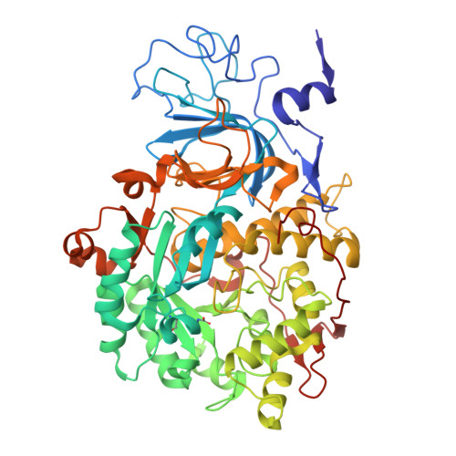
    8Q2E

  • PubMed Abstract: 

    This paper reports on the molecular details of the reactivity of urease, a nickel-dependent enzyme that catalyses the last step of organic nitrogen mineralization, with thiuram disulphides, a class of molecules known to inactivate the enzyme with high efficacy but for which the mechanism of action had not been yet established. IC 50 values of tetramethylthiuram disulphide (TMTD or Thiram) and tetraethylthiuram disulphide (TETD or Disulfiram) in the low micromolar range were determined for plant and bacterial ureases. The X-ray crystal structure of Sporosarcina pasteurii urease inactivated by Thiram, determined at 1.68 Å resolution, revealed the presence of a covalent modification of the catalytically essential cysteine residue. This is located on the flexible flap that modulates the size of the active site channel and cavity. Formation of a Cys-S-S-C(S)-N(CH 3 ) 2 functionality responsible for enzyme inactivation was observed. Quantum-mechanical calculations carried out to rationalise the large reactivity of the active site cysteine support the view that a conserved histidine residue, adjacent to the cysteine in the active site flap, modulates the charge and electron density along the thiol SH bond by shifting electrons towards the sulphur atom and rendering the thiol proton more reactive. We speculate that this proton could be transferred to the nickel-coordinated urea amide group to yield a molecule of ammonia from the generated C urea -NH 3 + functionality during catalysis.


  • Organizational Affiliation
    • Laboratory of Bioinorganic Chemistry, Department of Pharmacy and Biotechnology (FaBiT), University of Bologna, Viale Giuseppe Fanin 40, Bologna I-40127, Italy. Electronic address: luca.mazzei2@unibo.it.

Macromolecule Content 

  • Total Structure Weight: 89.34 kDa 
  • Atom Count: 7,051 
  • Modeled Residue Count: 792 
  • Deposited Residue Count: 792 
  • Unique protein chains: 3

Macromolecules

Find similar proteins by:|  3D Structure
Entity ID: 1
MoleculeChains  Sequence LengthOrganismDetailsImage
Urease subunit gamma100Sporosarcina pasteuriiMutation(s): 2 
EC: 3.5.1.5
UniProt
Find proteins for P41022 (Sporosarcina pasteurii)
Explore P41022 
Go to UniProtKB:  P41022
Entity Groups
Sequence Clusters30% Identity50% Identity70% Identity90% Identity95% Identity100% Identity
UniProt GroupP41022
Sequence Annotations
Expand
Reference Sequence
Find similar proteins by:|  3D Structure
Entity ID: 2
MoleculeChains  Sequence LengthOrganismDetailsImage
Urease subunit beta122Sporosarcina pasteuriiMutation(s): 0 
EC: 3.5.1.5
UniProt
Find proteins for P41021 (Sporosarcina pasteurii)
Explore P41021 
Go to UniProtKB:  P41021
Entity Groups
Sequence Clusters30% Identity50% Identity70% Identity90% Identity95% Identity100% Identity
UniProt GroupP41021
Sequence Annotations
Expand
Reference Sequence
Find similar proteins by:|  3D Structure
Entity ID: 3
MoleculeChains  Sequence LengthOrganismDetailsImage
Urease subunit alpha570Sporosarcina pasteuriiMutation(s): 0 
EC: 3.5.1.5
UniProt
Find proteins for P41020 (Sporosarcina pasteurii)
Explore P41020 
Go to UniProtKB:  P41020
Entity Groups
Sequence Clusters30% Identity50% Identity70% Identity90% Identity95% Identity100% Identity
UniProt GroupP41020
Sequence Annotations
Expand
Reference Sequence

Small Molecules

Ligands 5 Unique
IDChains Name / Formula / InChI Key2D Diagram3D Interactions
IS9
(Subject of Investigation/LOI)

Query on IS9


Download:Ideal Coordinates CCD File
HA [auth C]dimethylcarbamodithioic acid
C3 H7 N S2
MZGNSEAPZQGJRB-UHFFFAOYSA-N
SO4

Query on SO4


Download:Ideal Coordinates CCD File
J [auth A]
K [auth A]
L [auth A]
LA [auth C]
M [auth A]
J [auth A],
K [auth A],
L [auth A],
LA [auth C],
M [auth A],
MA [auth C],
NA [auth C],
OA [auth C],
PA [auth C],
Q [auth B],
QA [auth C],
R [auth B],
RA [auth C],
S [auth B],
T [auth B],
U [auth B]
SULFATE ION
O4 S
QAOWNCQODCNURD-UHFFFAOYSA-L
EDO

Query on EDO


Download:Ideal Coordinates CCD File
AA [auth C]
BA [auth C]
CA [auth C]
D [auth A]
DA [auth C]
AA [auth C],
BA [auth C],
CA [auth C],
D [auth A],
DA [auth C],
E [auth A],
EA [auth C],
F [auth A],
FA [auth C],
G [auth A],
GA [auth C],
H [auth A],
I [auth A],
N [auth B],
O [auth B],
P [auth B],
V [auth C],
W [auth C],
X [auth C],
Y [auth C],
Z [auth C]
1,2-ETHANEDIOL
C2 H6 O2
LYCAIKOWRPUZTN-UHFFFAOYSA-N
NI

Query on NI


Download:Ideal Coordinates CCD File
IA [auth C],
JA [auth C]
NICKEL (II) ION
Ni
VEQPNABPJHWNSG-UHFFFAOYSA-N
OH

Query on OH


Download:Ideal Coordinates CCD File
KA [auth C]HYDROXIDE ION
H O
XLYOFNOQVPJJNP-UHFFFAOYSA-M
Modified Residues  2 Unique
IDChains TypeFormula2D DiagramParent
CXM
Query on CXM
A
L-PEPTIDE LINKINGC6 H11 N O4 SMET
KCX
Query on KCX
C
L-PEPTIDE LINKINGC7 H14 N2 O4LYS

Experimental Data & Validation

Experimental Data

  • Method: X-RAY DIFFRACTION
  • Resolution: 1.68 Å
  • R-Value Free:  0.163 (Depositor), 0.174 (DCC) 
  • R-Value Work:  0.138 (Depositor), 0.153 (DCC) 
  • R-Value Observed: 0.139 (Depositor) 
Space Group: P 63 2 2
Unit Cell:
Length ( Å )Angle ( ˚ )
a = 131.511α = 90
b = 131.511β = 90
c = 189.017γ = 120
Software Package:
Software NamePurpose
REFMACrefinement
XDSdata reduction
Aimlessdata scaling
REFMACphasing

Structure Validation

View Full Validation Report


Ligand Structure Quality Assessment 


Entry History 

& Funding Information

Deposition Data


Funding OrganizationLocationGrant Number
University of BolognaItaly--

Revision History  (Full details and data files)

  • Version 1.0: 2023-11-01
    Type: Initial release
  • Version 1.1: 2023-11-15
    Changes: Data collection