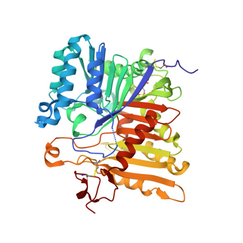
Structural analysis of PLD3 reveals insights into the mechanism of lysosomal 5' exonuclease-mediated nucleic acid degradation.
Roske, Y., Cappel, C., Cremer, N., Hoffmann, P., Koudelka, T., Tholey, A., Heinemann, U., Daumke, O., Damme, M.(2024) Nucleic Acids Res 52: 370-384
- PubMed: 37994783 Search on PubMedSearch on PubMed Central
- DOI: https://doi.org/10.1093/nar/gkad1114
- Primary Citation Related Structures:
8Q1K, 8Q1X - PubMed Abstract:
The phospholipase D (PLD) family is comprised of enzymes bearing phospholipase activity towards lipids or endo- and exonuclease activity towards nucleic acids. PLD3 is synthesized as a type II transmembrane protein and proteolytically cleaved in lysosomes, yielding a soluble active form. The deficiency of PLD3 leads to the slowed degradation of nucleic acids in lysosomes and chronic activation of nucleic acid-specific intracellular toll-like receptors. While the mechanism of PLD phospholipase activity has been extensively characterized, not much is known about how PLDs bind and hydrolyze nucleic acids. Here, we determined the high-resolution crystal structure of the luminal N-glycosylated domain of human PLD3 in its apo- and single-stranded DNA-bound forms. PLD3 has a typical phospholipase fold and forms homodimers with two independent catalytic centers via a newly identified dimerization interface. The structure of PLD3 in complex with an ssDNA-derived thymidine product in the catalytic center provides insights into the substrate binding mode of nucleic acids in the PLD family. Our structural data suggest a mechanism for substrate binding and nuclease activity in the PLD family and provide the structural basis to design immunomodulatory drugs targeting PLD3.
- Structural Biology, Max-Delbrück-Center for Molecular Medicine in the Helmholtz Association (MDC), 13125 Berlin, Germany.
Organizational Affiliation: