Flexible structural arrangement and DNA-binding properties of protein p6 from Bacillus subtillis phage phi 29.
Alcorlo, M., Luque-Ortega, J.R., Gago, F., Ortega, A., Castellanos, M., Chacon, P., de Vega, M., Blanco, L., Hermoso, J.M., Serrano, M., Rivas, G., Hermoso, J.A.(2024) Nucleic Acids Res 52: 2045-2065
- PubMed: 38281216 Search on PubMedSearch on PubMed Central
- DOI: https://doi.org/10.1093/nar/gkae041
- Primary Citation Related Structures:
8PW2, 8PW4 - PubMed Abstract:
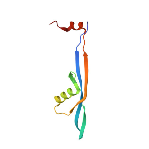
The genome-organizing protein p6 of Bacillus subtilis bacteriophage φ29 plays an essential role in viral development by activating the initiation of DNA replication and participating in the early-to-late transcriptional switch. These activities require the formation of a nucleoprotein complex in which the DNA adopts a right-handed superhelix wrapping around a multimeric p6 scaffold, restraining positive supercoiling and compacting the viral genome. Due to the absence of homologous structures, prior attempts to unveil p6's structural architecture failed. Here, we employed AlphaFold2 to engineer rational p6 constructs yielding crystals for three-dimensional structure determination. Our findings reveal a novel fold adopted by p6 that sheds light on its self-association mechanism and its interaction with DNA. By means of protein-DNA docking and molecular dynamic simulations, we have generated a comprehensive structural model for the nucleoprotein complex that consistently aligns with its established biochemical and thermodynamic parameters. Besides, through analytical ultracentrifugation, we have confirmed the hydrodynamic properties of the nucleocomplex, further validating in solution our proposed model. Importantly, the disclosed structure not only provides a highly accurate explanation for previously experimental data accumulated over decades, but also enhances our holistic understanding of the structural and functional attributes of protein p6 during φ29 infection.
- Department of Crystallography and Structural Biology, Institute of Physical-Chemistry "Blas Cabrera", CSIC, 28006 Madrid, Spain.
Organizational Affiliation: