8PUK | pdb_00008puk

ChiLob 7/4 H2 HC-T219C/C224S Kappa LC-E123C/C214S F(ab')2


Experimental Data Snapshot

  • Method: X-RAY DIFFRACTION
  • Resolution: 2.67 Å
  • R-Value Free: 
    0.282 (Depositor), 0.260 (DCC) 
  • R-Value Work: 
    0.242 (Depositor) 

Starting Model: experimental
View more details

wwPDB Validation 3D Report Full Report

Validation slider image for 8PUK

This is version 1.1 of the entry. See complete history

Literature

Structure-guided disulfide engineering restricts antibody conformation to elicit TNFR agonism.

Elliott, I.G.Fisher, H.Chan, H.T.C.Inzhelevskaya, T.Mockridge, C.I.Penfold, C.A.Duriez, P.J.Orr, C.M.Herniman, J.Muller, K.T.J.Essex, J.W.Cragg, M.S.Tews, I.

(2025) Nat Commun 16: 3495-3495

  • DOI: https://doi.org/10.1038/s41467-025-58773-8
  • Primary Citation Related Structures: 
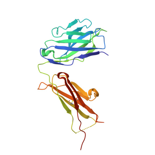
    8PUK, 8PUL

  • PubMed Abstract: 

    A promising strategy in cancer immunotherapy is activation of immune signalling pathways through antibodies that target co-stimulatory receptors. hIgG2, one of four human antibody isotypes, is known to deliver strong agonistic activity, and modification of hIgG2 hinge disulfides can influence immune-stimulating activity. This was shown for antibodies directed against the hCD40 receptor, where cysteine-to-serine exchange mutations caused changes in antibody conformational flexibility. Here we demonstrate that the principles of increasing agonism by restricting antibody conformation through disulfide modification can be translated to the co-stimulatory receptor h4-1BB, another member of the tumour necrosis factor receptor superfamily. Furthermore, we explore structure-guided design of the anti-hCD40 antibody ChiLob7/4 and show that engineering additional disulfides between opposing F(ab') arms can elicit conformational restriction, concomitant with enhanced agonism. These results support a mode where subtle increases in rigidity can deliver significant improvements in immunostimulatory activity, thus providing a strategy for the rational design of more powerful antibody therapeutics.


  • Organizational Affiliation
    • School of Chemistry and Chemical Engineering, Faculty of Engineering and Physical Sciences, University of Southampton, Southampton, SO17 1BJ, UK.

Macromolecule Content 

  • Total Structure Weight: 48.62 kDa 
  • Atom Count: 3,335 
  • Modeled Residue Count: 429 
  • Deposited Residue Count: 445 
  • Unique protein chains: 2

Macromolecules

Find similar proteins by:|  3D Structure
Entity ID: 1
MoleculeChains  Sequence LengthOrganismDetailsImage
Chilob 7/4 H2 heavy chain T219C/C224S231Homo sapiensMutation(s): 0 
Find similar proteins by:|  3D Structure
Entity ID: 2
MoleculeChains  Sequence LengthOrganismDetailsImage
Chilob 7/4 H2 kappa chain E123C/C214S214Homo sapiensMutation(s): 0 

Small Molecules

Ligands 1 Unique
IDChains Name / Formula / InChI Key2D Diagram3D Interactions
SO4

Query on SO4


Download:Ideal Coordinates CCD File
C [auth A]SULFATE ION
O4 S
QAOWNCQODCNURD-UHFFFAOYSA-L

Experimental Data & Validation

Experimental Data

  • Method: X-RAY DIFFRACTION
  • Resolution: 2.67 Å
  • R-Value Free:  0.282 (Depositor), 0.260 (DCC) 
  • R-Value Work:  0.242 (Depositor) 
Space Group: P 3 2 1
Unit Cell:
Length ( Å )Angle ( ˚ )
a = 148.167α = 90
b = 148.167β = 90
c = 45.295γ = 120
Software Package:
Software NamePurpose
REFMACrefinement
MxCuBEdata collection
XDSdata reduction
XSCALEdata scaling
MOLREPphasing
Cootmodel building
xia2data reduction

Structure Validation

View Full Validation Report


Entry History 

& Funding Information

Deposition Data


Funding OrganizationLocationGrant Number
Cancer Research UKUnited KingdomRCP010991

Revision History  (Full details and data files)

  • Version 1.0: 2025-03-05
    Type: Initial release
  • Version 1.1: 2025-07-16
    Changes: Database references