8PBP

Mutant R1785C of the dihydroorotase domain of human CAD protein bound to the substrate carbamoyl aspartate


Experimental Data Snapshot

  • Method: X-RAY DIFFRACTION
  • Resolution: 1.54 Å
  • R-Value Free: 0.194 
  • R-Value Work: 0.153 
  • R-Value Observed: 0.155 

wwPDB Validation   3D Report Full Report


Ligand Structure Quality Assessment 


This is version 1.2 of the entry. See complete history


Literature

Beyond genetics: Deciphering the impact of missense variants in CAD deficiency.

Del Cano-Ochoa, F.Ng, B.G.Rubio-Del-Campo, A.Mahajan, S.Wilson, M.P.Vilar, M.Rymen, D.Sanchez-Pintos, P.Kenny, J.Ley Martos, M.Campos, T.Wortmann, S.B.Freeze, H.H.Ramon-Maiques, S.

(2023) J Inherit Metab Dis 46: 1170-1185

  • DOI: https://doi.org/10.1002/jimd.12667
  • Primary Citation of Related Structures:  
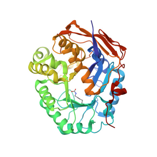
    8PBE, 8PBG, 8PBH, 8PBI, 8PBJ, 8PBK, 8PBM, 8PBN, 8PBP, 8PBQ, 8PBR, 8PBS, 8PBT, 8PBU

  • PubMed Abstract: 

    CAD is a large, 2225 amino acid multienzymatic protein required for de novo pyrimidine biosynthesis. Pathological CAD variants cause a developmental and epileptic encephalopathy which is highly responsive to uridine supplements. CAD deficiency is difficult to diagnose because symptoms are nonspecific, there is no biomarker, and the protein has over 1000 known variants. To improve diagnosis, we assessed the pathogenicity of 20 unreported missense CAD variants using a growth complementation assay that identified 11 pathogenic variants in seven affected individuals; they would benefit from uridine treatment. We also tested nine variants previously reported as pathogenic and confirmed the damaging effect of seven. However, we reclassified two variants as likely benign based on our assay, which is consistent with their long-term follow-up with uridine. We found that several computational methods are unreliable predictors of pathogenic CAD variants, so we extended the functional assay results by studying the impact of pathogenic variants at the protein level. We focused on CAD's dihydroorotase (DHO) domain because it accumulates the largest density of damaging missense changes. The atomic-resolution structures of eight DHO pathogenic variants, combined with functional and molecular dynamics analyses, provided a comprehensive structural and functional understanding of the activity, stability, and oligomerization of CAD's DHO domain. Combining our functional and protein structural analysis can help refine clinical diagnostic workflow for CAD variants in the genomics era.


  • Organizational Affiliation

    Structure of Macromolecular Targets Unit, Instituto de Biomedicina de Valencia (IBV), CSIC, Valencia, Spain.


Macromolecules
Find similar proteins by:  (by identity cutoff)  |  3D Structure
Entity ID: 1
MoleculeChains Sequence LengthOrganismDetailsImage
CAD protein362Homo sapiensMutation(s): 1 
Gene Names: CAD
EC: 6.3.5.5 (PDB Primary Data), 2.1.3.2 (PDB Primary Data), 3.5.2.3 (PDB Primary Data)
UniProt & NIH Common Fund Data Resources
Find proteins for P27708 (Homo sapiens)
Explore P27708 
Go to UniProtKB:  P27708
PHAROS:  P27708
GTEx:  ENSG00000084774 
Entity Groups  
Sequence Clusters30% Identity50% Identity70% Identity90% Identity95% Identity100% Identity
UniProt GroupP27708
Sequence Annotations
Expand
  • Reference Sequence
Small Molecules
Ligands 4 Unique
IDChains Name / Formula / InChI Key2D Diagram3D Interactions
NCD (Subject of Investigation/LOI)
Query on NCD

Download Ideal Coordinates CCD File 
B [auth A]N-CARBAMOYL-L-ASPARTATE
C5 H8 N2 O5
HLKXYZVTANABHZ-REOHCLBHSA-N
DOR (Subject of Investigation/LOI)
Query on DOR

Download Ideal Coordinates CCD File 
D [auth A](4S)-2,6-DIOXOHEXAHYDROPYRIMIDINE-4-CARBOXYLIC ACID
C5 H6 N2 O4
UFIVEPVSAGBUSI-REOHCLBHSA-N
ZN
Query on ZN

Download Ideal Coordinates CCD File 
F [auth A],
G [auth A],
H [auth A]
ZINC ION
Zn
PTFCDOFLOPIGGS-UHFFFAOYSA-N
FMT
Query on FMT

Download Ideal Coordinates CCD File 
C [auth A],
E [auth A]
FORMIC ACID
C H2 O2
BDAGIHXWWSANSR-UHFFFAOYSA-N
Modified Residues  1 Unique
IDChains TypeFormula2D DiagramParent
KCX
Query on KCX
A
L-PEPTIDE LINKINGC7 H14 N2 O4LYS
Experimental Data & Validation

Experimental Data

  • Method: X-RAY DIFFRACTION
  • Resolution: 1.54 Å
  • R-Value Free: 0.194 
  • R-Value Work: 0.153 
  • R-Value Observed: 0.155 
  • Space Group: C 2 2 21
Unit Cell:
Length ( Å )Angle ( ˚ )
a = 82.132α = 90
b = 158.552β = 90
c = 61.769γ = 90
Software Package:
Software NamePurpose
EDNAdata collection
autoPROCdata processing
PHASERphasing
REFMACrefinement
PHENIXrefinement

Structure Validation

View Full Validation Report


Ligand Structure Quality Assessment 


Entry History & Funding Information

Deposition Data


Funding OrganizationLocationGrant Number
Ministerio de Ciencia e Innovacion (MCIN)SpainPID2021-128468NB-I00
Other privateFundacion Ramon Areces, XX National Call
Other governmentPostdoctoral fellow of the Generalitat Valenciana (APOSTD 2021)

Revision History  (Full details and data files)

  • Version 1.0: 2023-11-01
    Type: Initial release
  • Version 1.1: 2023-11-15
    Changes: Data collection
  • Version 1.2: 2023-11-22
    Changes: Database references