8P3Y

Homomeric GluA2 flip R/G-edited Q/R-edited F231A mutant in tandem with TARP gamma-2, desensitized conformation 3


Experimental Data Snapshot

  • Method: ELECTRON MICROSCOPY
  • Resolution: 3.55 Å
  • Aggregation State: PARTICLE 
  • Reconstruction Method: SINGLE PARTICLE 

wwPDB Validation   3D Report Full Report


This is version 1.3 of the entry. See complete history


Literature

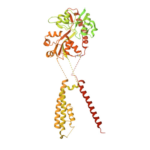
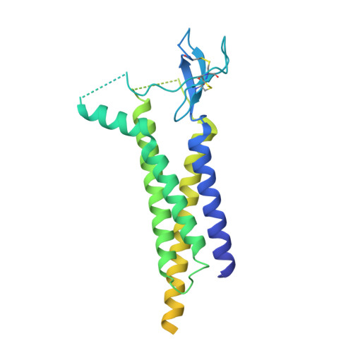
Structural mobility tunes signalling of the GluA1 AMPA glutamate receptor.

Zhang, D.Ivica, J.Krieger, J.M.Ho, H.Yamashita, K.Stockwell, I.Baradaran, R.Cais, O.Greger, I.H.

(2023) Nature 621: 877-882

  • DOI: https://doi.org/10.1038/s41586-023-06528-0
  • Primary Citation of Related Structures:  
    8C1P, 8C1Q, 8C1R, 8C1S, 8C2H, 8C2I, 8P3Q, 8P3S, 8P3T, 8P3U, 8P3V, 8P3W, 8P3X, 8P3Y, 8P3Z, 8PIV

  • PubMed Abstract: 

    AMPA glutamate receptors (AMPARs), the primary mediators of excitatory neurotransmission in the brain, are either GluA2 subunit-containing and thus Ca 2+ -impermeable, or GluA2-lacking and Ca 2+ -permeable 1 . Despite their prominent expression throughout interneurons and glia, their role in long-term potentiation and their involvement in a range of neuropathologies 2 , structural information for GluA2-lacking receptors is currently absent. Here we determine and characterize cryo-electron microscopy structures of the GluA1 homotetramer, fully occupied with TARPγ3 auxiliary subunits (GluA1/γ3). The gating core of both resting and open-state GluA1/γ3 closely resembles GluA2-containing receptors. However, the sequence-diverse N-terminal domains (NTDs) give rise to a highly mobile assembly, enabling domain swapping and subunit re-alignments in the ligand-binding domain tier that are pronounced in desensitized states. These transitions underlie the unique kinetic properties of GluA1. A GluA2 mutant (F231A) increasing NTD dynamics phenocopies this behaviour, and exhibits reduced synaptic responses, reflecting the anchoring function of the AMPAR NTD at the synapse. Together, this work underscores how the subunit-diverse NTDs determine subunit arrangement, gating properties and ultimately synaptic signalling efficiency among AMPAR subtypes.


  • Organizational Affiliation

    Neurobiology Division, Medical Research Council (MRC) Laboratory of Molecular Biology, Cambridge, UK.


Macromolecules
Find similar proteins by:  (by identity cutoff)  |  3D Structure
Entity ID: 1
MoleculeChains Sequence LengthOrganismDetailsImage
Glutamate receptor 2
A, B, C, D
881Rattus norvegicusMutation(s): 1 
Gene Names: Gria2Glur2
Membrane Entity: Yes 
UniProt
Find proteins for P19491 (Rattus norvegicus)
Explore P19491 
Go to UniProtKB:  P19491
Entity Groups  
Sequence Clusters30% Identity50% Identity70% Identity90% Identity95% Identity100% Identity
UniProt GroupP19491
Sequence Annotations
Expand
  • Reference Sequence
Find similar proteins by:  (by identity cutoff)  |  3D Structure
Entity ID: 2
MoleculeChains Sequence LengthOrganismDetailsImage
Voltage-dependent calcium channel gamma-2 subunit
E, F, G, H
323Rattus norvegicusMutation(s): 0 
Gene Names: Cacng2Stg
Membrane Entity: Yes 
UniProt
Find proteins for Q71RJ2 (Rattus norvegicus)
Explore Q71RJ2 
Go to UniProtKB:  Q71RJ2
Entity Groups  
Sequence Clusters30% Identity50% Identity70% Identity90% Identity95% Identity100% Identity
UniProt GroupQ71RJ2
Sequence Annotations
Expand
  • Reference Sequence
Experimental Data & Validation

Experimental Data

  • Method: ELECTRON MICROSCOPY
  • Resolution: 3.55 Å
  • Aggregation State: PARTICLE 
  • Reconstruction Method: SINGLE PARTICLE 
EM Software:
TaskSoftware PackageVersion
MODEL REFINEMENTCoot0.9.8.4-pre

Structure Validation

View Full Validation Report


Entry History & Funding Information

Deposition Data


Funding OrganizationLocationGrant Number
Medical Research Council (MRC, United Kingdom)United KingdomMC_U105174197
H2020 Marie Curie Actions of the European CommissionEuropean Union101024130
Wellcome TrustUnited Kingdom223194/Z/21/Z

Revision History  (Full details and data files)

  • Version 1.0: 2023-08-30
    Type: Initial release
  • Version 1.1: 2023-09-13
    Changes: Database references
  • Version 1.2: 2023-09-20
    Changes: Database references
  • Version 1.3: 2023-10-11
    Changes: Database references