8P2Q

Crystal structure of Hen Egg White Lysozyme co-crystallized with 10 mM TbXo4-OH


Experimental Data Snapshot

  • Method: X-RAY DIFFRACTION
  • Resolution: 1.69 Å
  • R-Value Free: 0.195 
  • R-Value Work: 0.159 
  • R-Value Observed: 0.161 

Starting Model: experimental
View more details

wwPDB Validation   3D Report Full Report


Ligand Structure Quality Assessment 


This is version 1.2 of the entry. See complete history


Literature

Influence of Chemical Modifications of the Crystallophore on Protein Nucleating Properties and Supramolecular Interactions Network.

Roux, A.Alsalman, Z.Jiang, T.Mulatier, J.C.Pitrat, D.Dumont, E.Riobe, F.Gillet, N.Girard, E.Maury, O.

(2024) Chemistry 30: e202400900-e202400900

  • DOI: https://doi.org/10.1002/chem.202400900
  • Primary Citation of Related Structures:  
    8OWC, 8P2Q, 8PIW, 8POB

  • PubMed Abstract: 

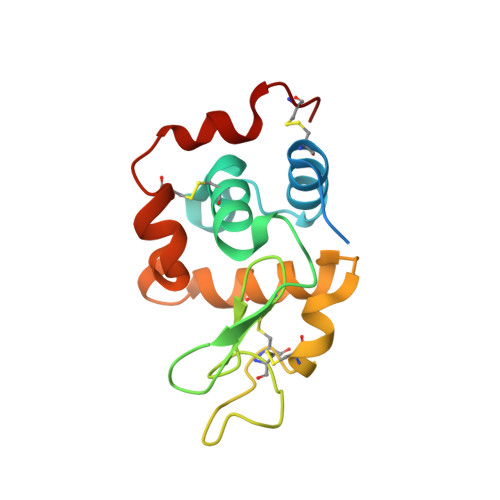
    Crystallophores are lanthanide complexes that have demonstrated outstanding induction of crystallization for various proteins. This article explores the effect of tailored modifications of the crystallophore first generation and their impact on the nucleating properties and protein crystal structures. Through high-throughput crystallization experiments and dataset analysis, we evaluated the effectiveness of these variants, in comparison to the first crystallophore generation G 1 . In particular, the V 1 variant, featuring a propanol pendant arm, demonstrated the ability to produce new crystallization conditions for the proteins tested (hen-egg white lysozyme, proteinase K and thaumatin). Structural analysis performed in the case of hen egg-white lysozyme along with Molecular Dynamics simulations, highlights V 1 's unique behavior, taking advantage of the flexibility of its propanol arm to explore different protein surfaces and form versatile supramolecular interactions.


  • Organizational Affiliation

    Univ. Lyon, École Normale Supérieure de Lyon, CNRS UMR 5182, Laboratoire de Chimie, 46 allée d'Italie, 69007, Lyon, France.


Macromolecules
Find similar proteins by:  (by identity cutoff)  |  3D Structure
Entity ID: 1
MoleculeChains Sequence LengthOrganismDetailsImage
Lysozyme C147Gallus gallusMutation(s): 0 
EC: 3.2.1.17
UniProt
Find proteins for P00698 (Gallus gallus)
Explore P00698 
Go to UniProtKB:  P00698
Entity Groups  
Sequence Clusters30% Identity50% Identity70% Identity90% Identity95% Identity100% Identity
UniProt GroupP00698
Sequence Annotations
Expand
  • Reference Sequence
Experimental Data & Validation

Experimental Data

  • Method: X-RAY DIFFRACTION
  • Resolution: 1.69 Å
  • R-Value Free: 0.195 
  • R-Value Work: 0.159 
  • R-Value Observed: 0.161 
  • Space Group: P 43 21 2
Unit Cell:
Length ( Å )Angle ( ˚ )
a = 77.143α = 90
b = 77.143β = 90
c = 37.695γ = 90
Software Package:
Software NamePurpose
PHENIXrefinement
XDSdata reduction
SCALAdata scaling
PHASERphasing

Structure Validation

View Full Validation Report


Ligand Structure Quality Assessment 


Entry History & Funding Information

Deposition Data


Funding OrganizationLocationGrant Number
Other governmentFranceProject XO4_2.0

Revision History  (Full details and data files)

  • Version 1.0: 2024-06-05
    Type: Initial release
  • Version 1.1: 2024-07-10
    Changes: Database references
  • Version 1.2: 2024-07-17
    Changes: Database references