8P15 | pdb_00008p15

Cryo-EM structure of Rhodopsin-Gi bound with antibody fragments scFv16 and Fab79, conformation 2


Experimental Data Snapshot

  • Method: ELECTRON MICROSCOPY
  • Resolution: 5.90 Å
  • Aggregation State: PARTICLE 
  • Reconstruction Method: SINGLE PARTICLE 

Starting Models: experimental, in silico
View more details

wwPDB Validation   3D Report Full Report


This is version 1.1 of the entry. See complete history


Literature

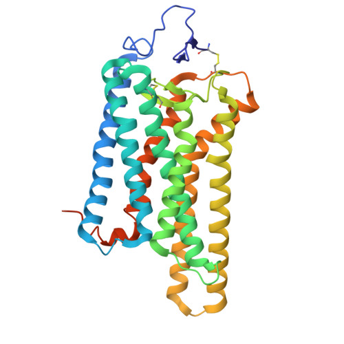
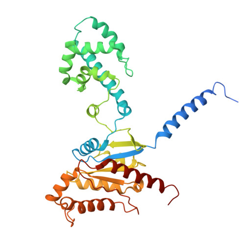
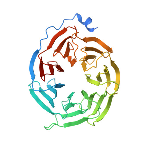
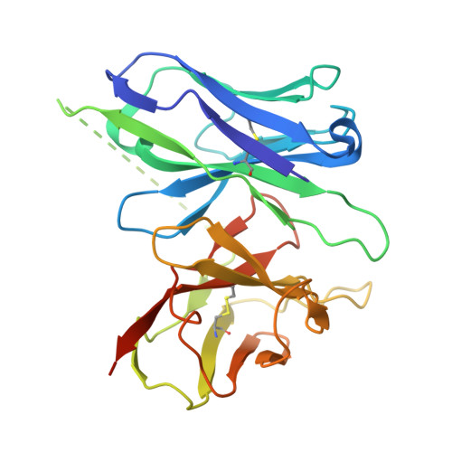
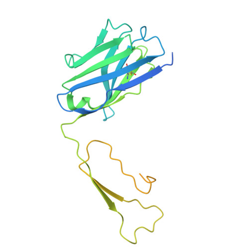
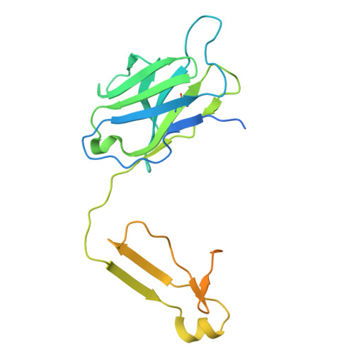
Tool antibody fragments reveal multiple conformations of the rhodopsin-Gi signaling complex.

Pamula, F.Tejero, O.Muhle, J.Thoma, R.Schertler, G.F.X.Marino, J.Tsai, C.J.

(2025) Biophys J 

  • DOI: https://doi.org/10.1016/j.bpj.2025.09.044
  • Primary Citation of Related Structures:  
    8P12, 8P13, 8P15

  • PubMed Abstract: 

    Antibody Fab fragments are widely used protein binders that assist in structural studies of G-protein-coupled receptor (GPCR) signaling complexes. Expanding the repertoire of such binders to target distinct components of the signaling complex offers opportunities to probe conformational regulation and dynamics. Here, we report the biochemical and cryo-EM characterization of two Fab fragments, Fab79 and Fab13, raised against the rhodopsin-Gαiβγ complex. Fab79 binds to the flexible α-helical domain (AHD) of the Gαi subunit and prevents complex dissociation in the presence of the nonhydrolyzable GTP analog, GTPγS, likely by hindering AHD closure, a step necessary for complex dissociation. In contrast, Fab13 binds rigidly to Gβ without directly contacting Gα or the receptor. These findings show that Fab79 and Fab13 reveal functionally relevant conformational states of G-protein activation and serve as practical tools to stabilize or modulate GPCR signaling complexes.


  • Organizational Affiliation
    • Laboratory of Biomolecular Research, Paul Scherrer Institute, Villigen, Switzerland; Department of Biology, ETH Zürich, Zürich, Switzerland.

Macromolecules
Find similar proteins by:  (by identity cutoff)  |  3D Structure
Entity ID: 1
MoleculeChains Sequence LengthOrganismDetailsImage
RhodopsinA [auth R]348Bos taurusMutation(s): 3 
Gene Names: RHO
UniProt
Find proteins for P02699 (Bos taurus)
Explore P02699 
Go to UniProtKB:  P02699
Entity Groups  
Sequence Clusters30% Identity50% Identity70% Identity90% Identity95% Identity100% Identity
UniProt GroupP02699
Sequence Annotations
Expand
  • Reference Sequence
Find similar proteins by:  (by identity cutoff)  |  3D Structure
Entity ID: 2
MoleculeChains Sequence LengthOrganismDetailsImage
Guanine nucleotide-binding protein G(i) subunit alpha-1B [auth A]376Homo sapiensMutation(s): 0 
Gene Names: GNAI1
EC: 3.6.5
UniProt & NIH Common Fund Data Resources
Find proteins for P63096 (Homo sapiens)
Explore P63096 
Go to UniProtKB:  P63096
PHAROS:  P63096
GTEx:  ENSG00000127955 
Entity Groups  
Sequence Clusters30% Identity50% Identity70% Identity90% Identity95% Identity100% Identity
UniProt GroupP63096
Sequence Annotations
Expand
  • Reference Sequence
Find similar proteins by:  (by identity cutoff)  |  3D Structure
Entity ID: 3
MoleculeChains Sequence LengthOrganismDetailsImage
Guanine nucleotide-binding protein G(I)/G(S)/G(T) subunit beta-1C [auth B]340Bos taurusMutation(s): 0 
UniProt
Find proteins for P62871 (Bos taurus)
Explore P62871 
Go to UniProtKB:  P62871
Entity Groups  
Sequence Clusters30% Identity50% Identity70% Identity90% Identity95% Identity100% Identity
UniProt GroupP62871
Sequence Annotations
Expand
  • Reference Sequence
Find similar proteins by:  (by identity cutoff)  |  3D Structure
Entity ID: 4
MoleculeChains Sequence LengthOrganismDetailsImage
Guanine nucleotide-binding protein G(T) subunit gamma-T1D [auth G]74Bos taurusMutation(s): 0 
UniProt
Find proteins for P02698 (Bos taurus)
Explore P02698 
Go to UniProtKB:  P02698
Entity Groups  
Sequence Clusters30% Identity50% Identity70% Identity90% Identity95% Identity100% Identity
UniProt GroupP02698
Sequence Annotations
Expand
  • Reference Sequence
Find similar proteins by:  (by identity cutoff)  |  3D Structure
Entity ID: 5
MoleculeChains Sequence LengthOrganismDetailsImage
single-chain Fv scFv16E [auth S]259Mus musculusMutation(s): 0 
Entity Groups  
Sequence Clusters30% Identity50% Identity70% Identity90% Identity95% Identity100% Identity
Sequence Annotations
Expand
  • Reference Sequence
Find similar proteins by:  (by identity cutoff)  |  3D Structure
Entity ID: 6
MoleculeChains Sequence LengthOrganismDetailsImage
Immunoglobin G heavy chain FAB fragmentF [auth H]262Mus musculusMutation(s): 0 
Entity Groups  
Sequence Clusters30% Identity50% Identity70% Identity90% Identity95% Identity100% Identity
Sequence Annotations
Expand
  • Reference Sequence
Find similar proteins by:  (by identity cutoff)  |  3D Structure
Entity ID: 7
MoleculeChains Sequence LengthOrganismDetailsImage
Immunoglobin G light chainG [auth L]239Mus musculusMutation(s): 0 
Entity Groups  
Sequence Clusters30% Identity50% Identity70% Identity90% Identity95% Identity100% Identity
Sequence Annotations
Expand
  • Reference Sequence
Experimental Data & Validation

Experimental Data

  • Method: ELECTRON MICROSCOPY
  • Resolution: 5.90 Å
  • Aggregation State: PARTICLE 
  • Reconstruction Method: SINGLE PARTICLE 
EM Software:
TaskSoftware PackageVersion
MODEL REFINEMENTPHENIX1.20.1-4487
RECONSTRUCTIONRELION3.1

Structure Validation

View Full Validation Report


Entry History & Funding Information

Deposition Data


Funding OrganizationLocationGrant Number
Swiss National Science FoundationSwitzerland173335
Swiss National Science FoundationSwitzerland153145
Swiss National Science FoundationSwitzerland192760
European Research Council (ERC)European Union951644

Revision History  (Full details and data files)

  • Version 1.0: 2024-11-20
    Type: Initial release
  • Version 1.1: 2025-10-29
    Changes: Data collection, Database references